Copyright
©The Author(s) 2025.
World J Gastrointest Endosc. Aug 16, 2025; 17(8): 111141
Published online Aug 16, 2025. doi: 10.4253/wjge.v17.i8.111141
Published online Aug 16, 2025. doi: 10.4253/wjge.v17.i8.111141
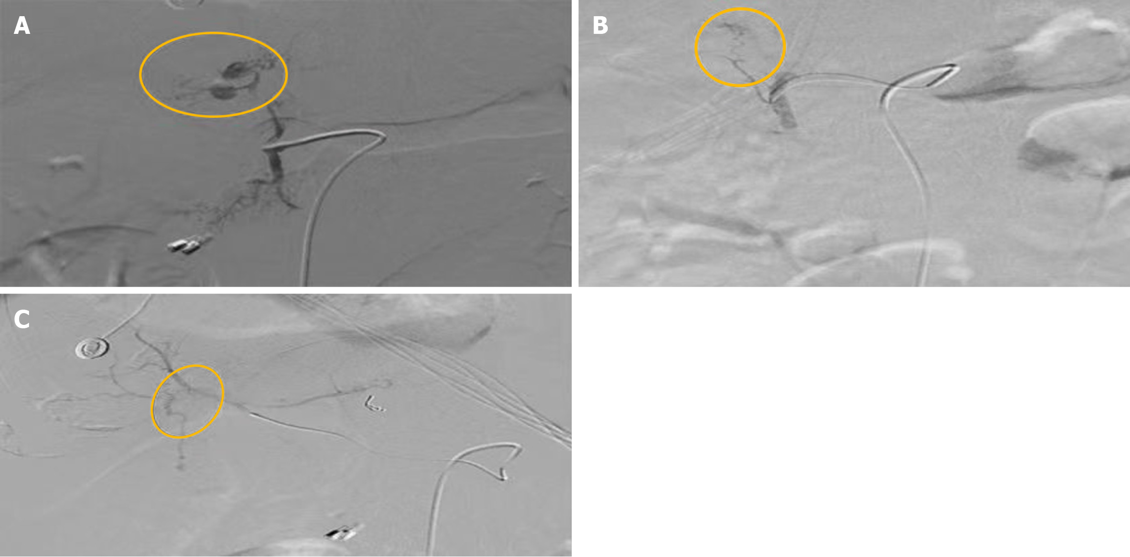
Figure 1 Bleeding part.
A: Bleeding from a small branch of the gastroduodenal artery; B: Bleeding from a small arterial branch of the gastroduodenal artery supplying the left lobe of the liver; C: Bleeding from a small arterial branch of the right hepatic artery anastomosing with the left hepatic artery.
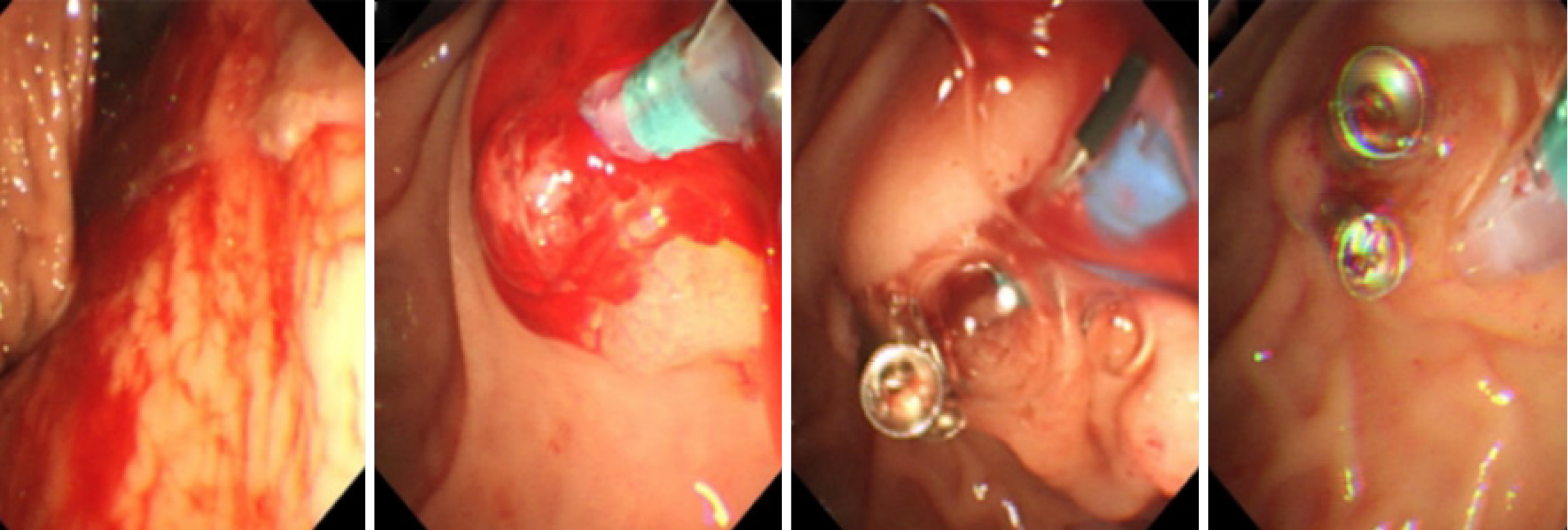
Figure 2
The entire process of endoscopic hemostasis.
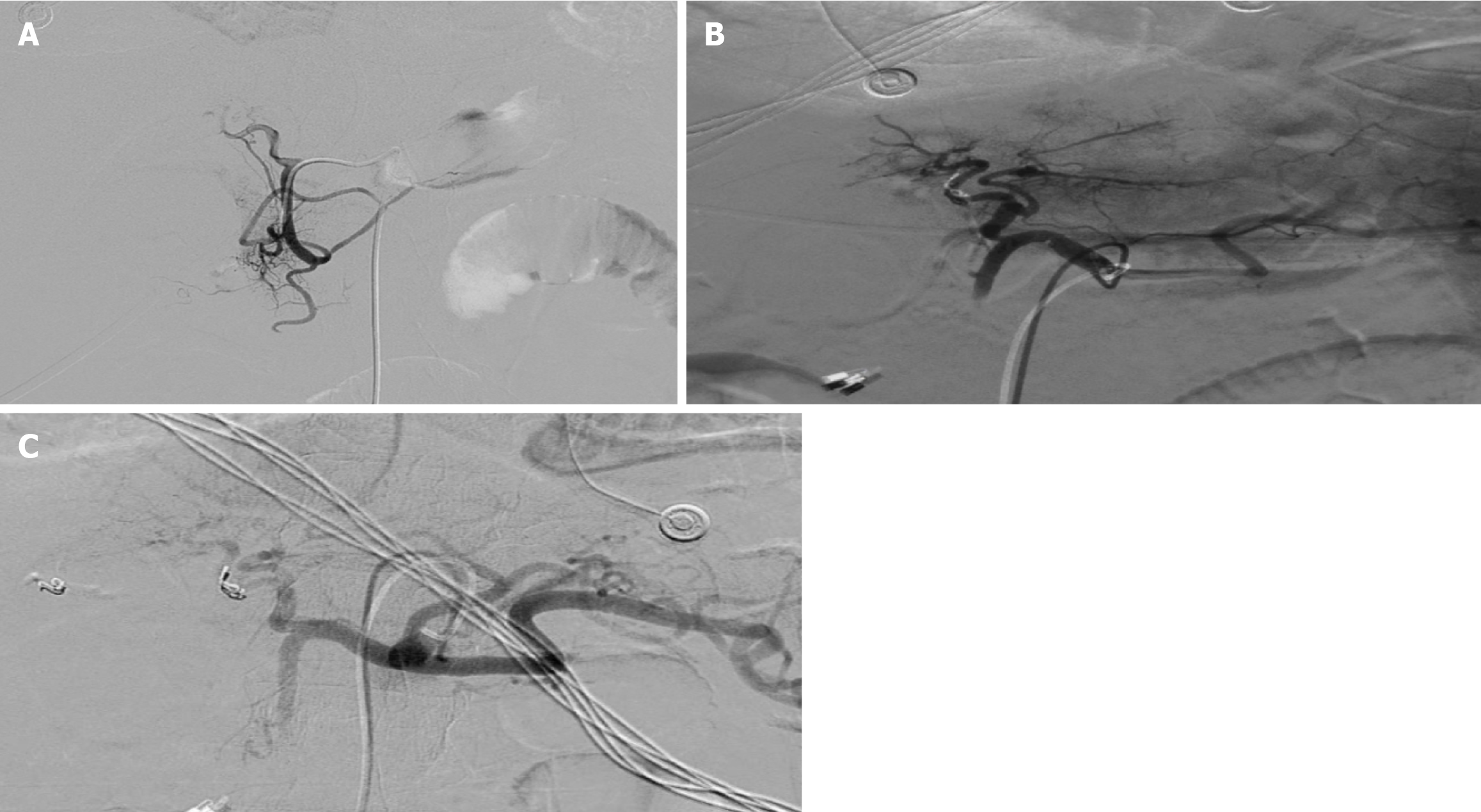
Figure 3 Disappearance of the bleeding point.
A: After interventional embolization, the bleeding points in the small branches of the gastroduodenal artery were no longer detectable; B: After interventional embolization, the bleeding points in the small arterial branch of the gastroduodenal artery supplying the left lobe of the liver were no longer detectable; C: After interventional embolization, the bleeding points in the small arterial branch of the right hepatic artery anastomosing with the left hepatic artery were no longer detectable.
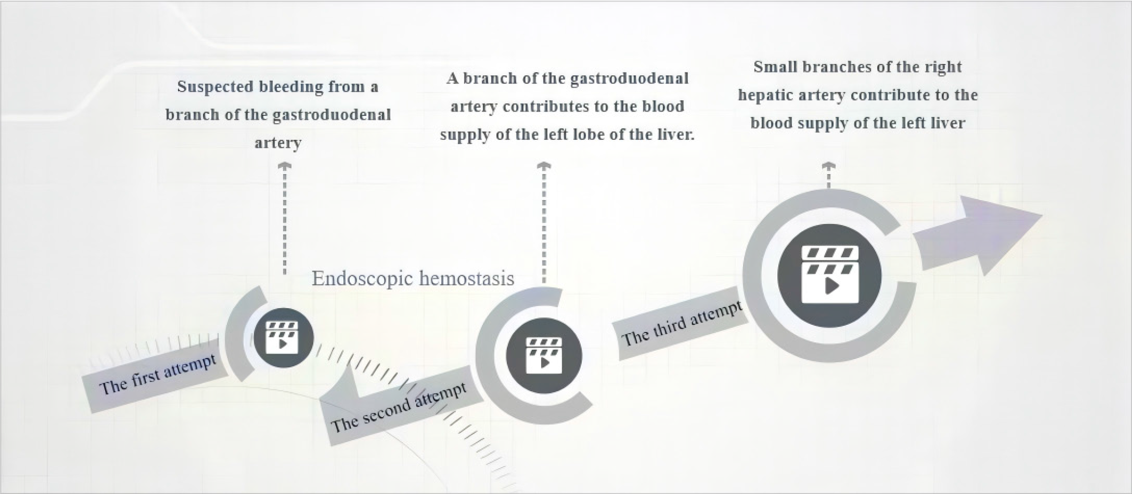
Figure 4
Management flowchart for delayed post-endoscopic retrograde cholangiopancreatography bleeding.
- Citation: Ma ZW, Gong XJ, Chen YJ, Wang B. Vascular anomaly as a cause of late bleeding after endoscopic retrograde cholangiopancreatography: A case report. World J Gastrointest Endosc 2025; 17(8): 111141
- URL: https://www.wjgnet.com/1948-5190/full/v17/i8/111141.htm
- DOI: https://dx.doi.org/10.4253/wjge.v17.i8.111141


