Copyright
©The Author(s) 2025.
World J Gastrointest Endosc. Aug 16, 2025; 17(8): 109104
Published online Aug 16, 2025. doi: 10.4253/wjge.v17.i8.109104
Published online Aug 16, 2025. doi: 10.4253/wjge.v17.i8.109104
Figure 1 Endoscopic view of the fistulas of Patient 1.
A: Lower fistulous tract with a channel; B: Openings of the upper fistulous tract above the anastomosis; C: Result after 10 days of therapy, with a residual cavity formed.
Figure 2 PandiCath®.
1: Channel for inflating balloons 5 and 8; 2: Main channel for fluid infusion, passive drainage, and shunting of the proximal and distal sections of the organ; 3: Manipulation channel connected to a controlled negative pressure pump, ensuring outflow from the isolated area or administration of medications; 4: Perforation openings above the isolated area; 5 and 8: Inflatable isolation balloons positioned above and below the defect area; 6: Opening of the manipulation channel (3), located between the balloons, allowing for biological material collection; 7: Protrusions around opening 6, preventing adhesion of the mucous layer; 9: Distal opening of channel 2.
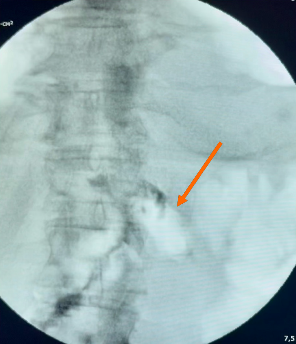
Figure 3 Fluoroscopy of the anastomosis area three days after the start of therapy of Patient 2.
The arrow indicates traces of contrast medium and air in the jejunal stump anastomosed according to Roux, with no contrast leakage detected.
- Citation: Kashintsev AA, Eselevich RV, Surov DA, Balyura OV, Proutski V. Isolation techniques for gastrointestinal tract defects: Two case reports. World J Gastrointest Endosc 2025; 17(8): 109104
- URL: https://www.wjgnet.com/1948-5190/full/v17/i8/109104.htm
- DOI: https://dx.doi.org/10.4253/wjge.v17.i8.109104


