©The Author(s) 2025.
World J Gastrointest Endosc. Jul 16, 2025; 17(7): 108082
Published online Jul 16, 2025. doi: 10.4253/wjge.v17.i7.108082
Published online Jul 16, 2025. doi: 10.4253/wjge.v17.i7.108082
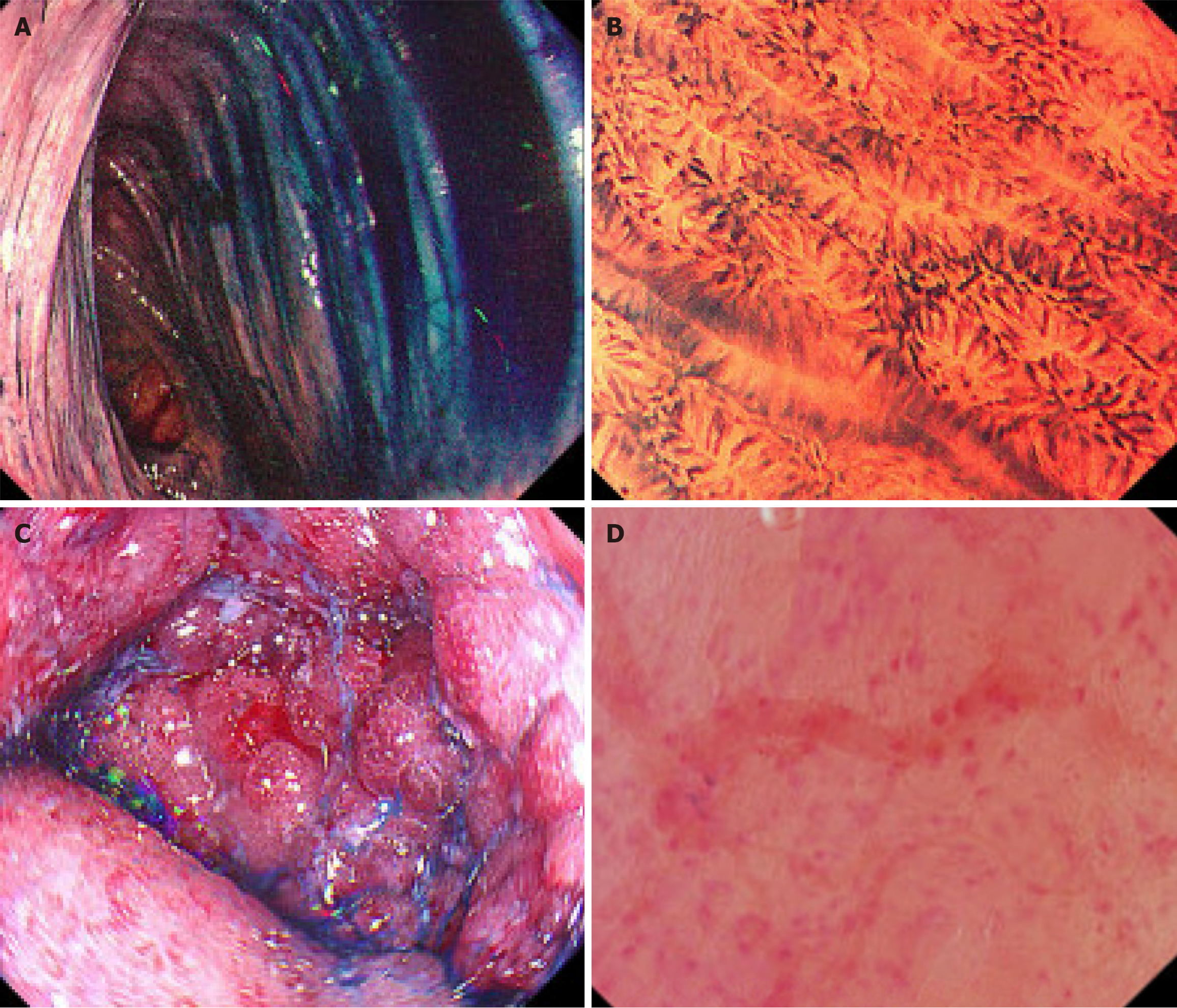
Figure 1 Corresponding endoscopic and endocytoscopic evaluations.
A: Normal vascular pattern of the colonic mucosa, graded as Mayo endoscopic score 0; B: Endocytoscopy score 0 for the same patient as A, characterized by normal crypt shape (0), crypt distance (0), and visible small vessels (0). The Erlangen Endocytoscopy in ColiTis score is 0, including normal crypt shape (0), crypt distance (0), and normal vascular structure (0), absence inflammatory cell infiltrate (0), and absence of crypt abscess (0). This is graded as Nancy histological index 0, indicating histological remission; C: Marked erythematous mucosa with an absent vascular pattern, friability, and erosions, graded as Mayo endoscopic score 2; D: Endocytoscopy score 4 for the same patient as C, characterized by irregular crypt shape (2), intermediate crypt distance (1), and visible vessels (1). The ErLangen Endocytoscopy in ColiTis score is 4, including irregular crypt shape (1), increased distance between crypts (1), distorted vascular structure (1), presence of inflammatory cell infiltrate (1), and absence of crypt abscess (0). This is graded as Nancy histological index 3, indicating the presence of moderate to severe chronic and acute inflammatory cells.
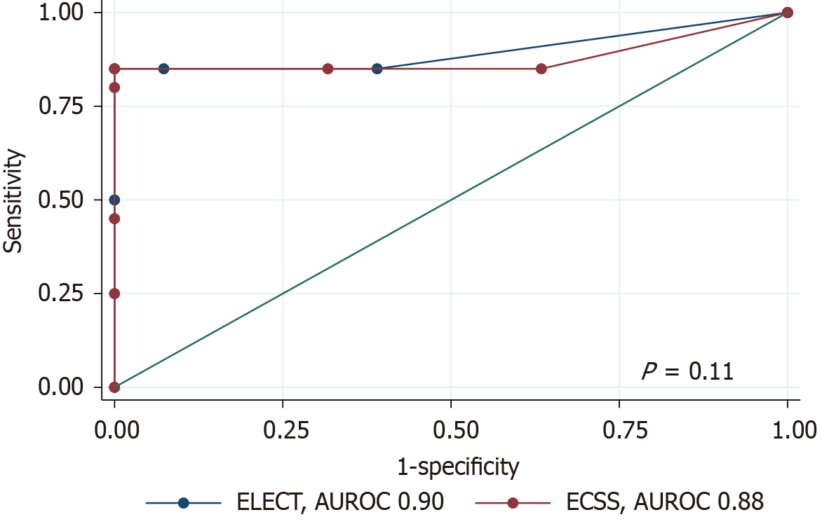
Figure 2 Comparison of diagnostic performance between endocytoscopy score and the Erlangen Endocytoscopy in ColiTis score.
ELECT: ErLangen Endocytoscopy in ColiTis; AUROC: Area under the receiver operating characteristic curve; ECSS: Endocytoscopy score.
- Citation: Chaemsupaphan T, Shir Ali M, Fung C, Paramsothy S, Leong RW. Endocytoscopy in real-time assessment of histological and endoscopic activity in ulcerative colitis. World J Gastrointest Endosc 2025; 17(7): 108082
- URL: https://www.wjgnet.com/1948-5190/full/v17/i7/108082.htm
- DOI: https://dx.doi.org/10.4253/wjge.v17.i7.108082


