©The Author(s) 2025.
World J Gastrointest Endosc. Jul 16, 2025; 17(7): 107911
Published online Jul 16, 2025. doi: 10.4253/wjge.v17.i7.107911
Published online Jul 16, 2025. doi: 10.4253/wjge.v17.i7.107911
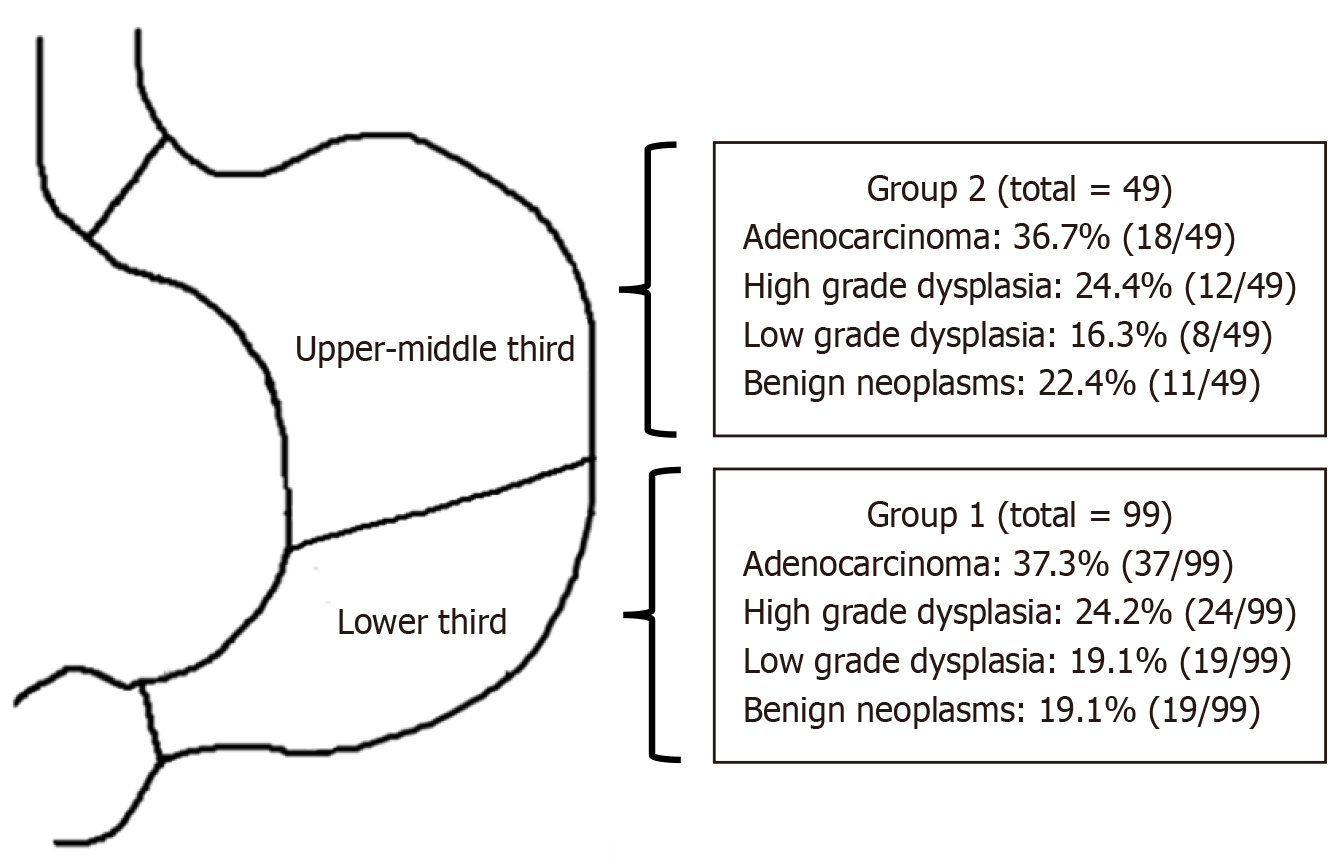
Figure 1
Schematic of the location of stomach lesions.
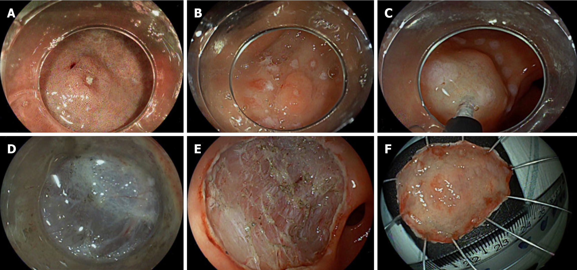
Figure 2 Illustrative pictures of endoscopic submucosal dissection in the lower third of the stomach.
A: A 33-year-old woman presented with an elevated-type lesion (0-IIa) in the lower third of the stomach (gastric antrum); B: Markings were placed for endoscopic submucosal dissection, mucosal incision was started at the distal margin of the lesion; C: Submucosal injection to lift the lesion with 0.4% sodium hyaluronate in a teardrops form; D: Submucosal layer dissection; E: Complete tumor resection was achieved en bloc; F: Histological analysis of the endoscopic submucosal dissection specimen that revealed high grade dysplasia.
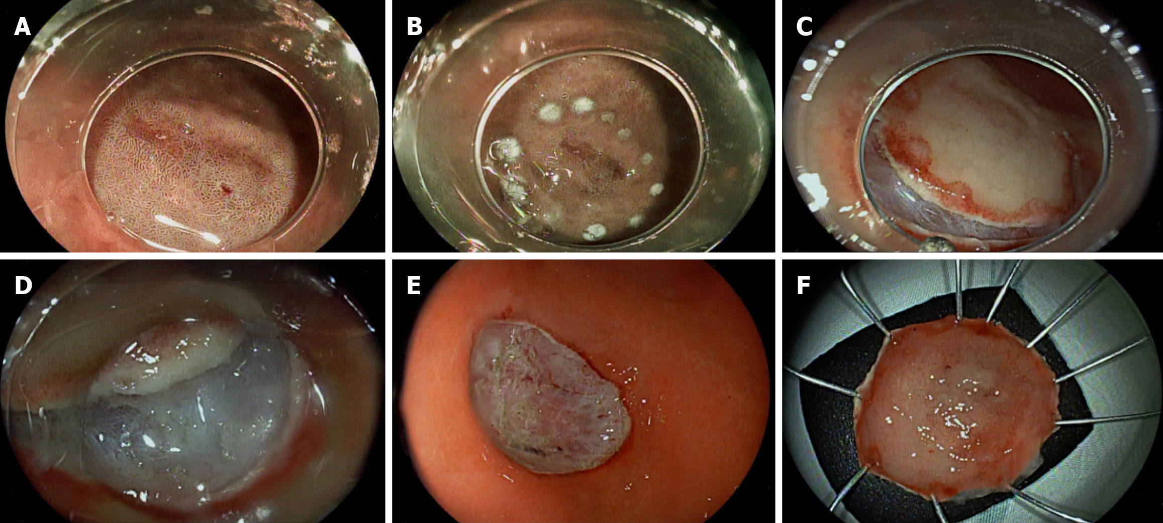
Figure 3 Illustrative pictures of endoscopic submucosal dissection in the upper-middle third of the stomach.
A: A 33-year-old woman presented with a flat-type lesion (0-IIb) in the upper-middle third of the stomach (gastric body); B: Markings were placed for endoscopic submucosal dissection. Mucosal incision was started at the distal margin of the lesion; C: Mucosal incision; D: Submucosal layer dissection; E: Complete tumor resection was achieved en bloc; F: Histological analysis of the endoscopic submucosal dissection specimen that revealed predominantly undifferentiated (diffuse) mixed-type adenocarcinoma.
- Citation: Aliaga Ramos J, Arantes VN. Impact of gastric neoplasms location on clinical outcome of patients treated by endoscopic submucosal dissection. World J Gastrointest Endosc 2025; 17(7): 107911
- URL: https://www.wjgnet.com/1948-5190/full/v17/i7/107911.htm
- DOI: https://dx.doi.org/10.4253/wjge.v17.i7.107911


