©The Author(s) 2025.
World J Gastrointest Endosc. Jul 16, 2025; 17(7): 107872
Published online Jul 16, 2025. doi: 10.4253/wjge.v17.i7.107872
Published online Jul 16, 2025. doi: 10.4253/wjge.v17.i7.107872
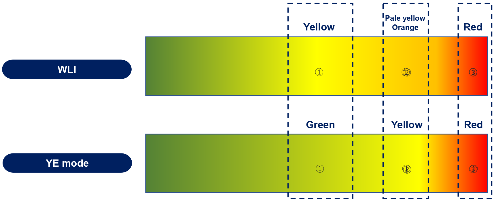
Figure 1 Changes in colour between white light mode and yellow enhancement mode done by yellow enhancement's image processor[14].
Image obtained from Olympus corporation. YE: Yellow enhancement; WLI: White light imaging. Citation: Singh H, Suherman R, Koh FH. Yellow enhancement imaging facilitates identification of surgical planes and key structures during challenging high anterior resection in a patient with obesity. Ann Coloproctol 2025. Copyright© The Authors 2025. Published by The Korean Society of Coloproctology Publishers. The authors have obtained the permission (Supplementary material).
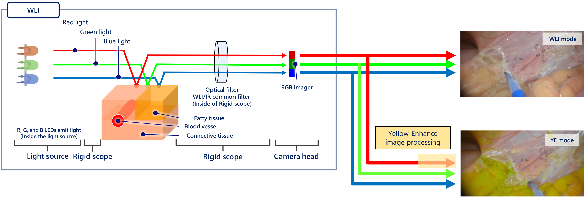
Figure 2 The process by which yellow enhancement converts white light images into yellow enhanced images[14].
Citation: Singh H, Suherman R, Koh FH. Yellow enhancement imaging facilitates identification of surgical planes and key structures during challenging high anterior resection in a patient with obesity. Ann Coloproctol 2025. Copyright© The Authors 2025. Published by The Korean Society of Coloproctology Publishers. The authors have obtained the permission (Supplementary material).
Figure 3 Surgical planes with white light (left) and yellow enhancement (right)[14].
Avascular segments are easier to identify with YE and allow for safer dissections as indicated by the dotted lines. A: Left; B: Right. Citation: Singh H, Suherman R, Koh FH. Yellow enhancement imaging facilitates identification of surgical planes and key structures during challenging high anterior resection in a patient with obesity. Ann Coloproctol 2025. Copyright© The Authors 2025. Published by The Korean Society of Coloproctology Publishers. The authors have obtained the permission (Supplementary material).
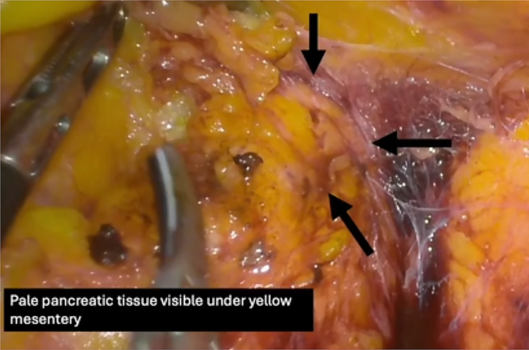
Figure 4 Identification of Pancreatic tissue underneath peripancreatic and mesenteric fat tissue[13].
Citation: Suherman RC, Singh H, Aw DKL, Chong CXZ, Ng JL, Sivarajah S, Tan WJ, Foo FJ, Ladlad J, Khoo N, Tan CHM, Koh FH. The role of yellow enhancement in laparoscopy. Br J Surg 2024; 111. Copyright© The Authors 2024. Published by Oxford University Press on behalf of BJS Foundation Ltd. The authors have obtained the permission (Supplementary material).
Figure 5 Usage of yellow enhancement in tracing and clipping of the inferior mesenteric artery in a laparoscopic anterior resection[14].
A: Demonstration of the skeletonised inferior mesenteric artery amidst a lack of space from encroaching bowel in an obese patient with short colonic mesentery; B: Application of a hemostatic clip on the inferior mesenteric artery. Citation: Singh H, Suherman R, Koh FH. Yellow enhancement imaging facilitates identification of surgical planes and key structures during challenging high anterior resection in a patient with obesity. Ann Coloproctol 2025. Copyright© The Authors 2025. Published by The Korean Society of Coloproctology Publishers. The authors have obtained the permission (Supplementary material).
Figure 6 Hypogastric nerve being identified and, in this case, sacrificed[14].
Citation: Singh H, Suherman R, Koh FH. Yellow enhancement imaging facilitates identification of surgical planes and key structures during challenging high anterior resection in a patient with obesity. Ann Coloproctol 2025. Copyright© The Authors 2025. Published by The Korean Society of Coloproctology Publishers. The authors have obtained the permission (Supplementary material).
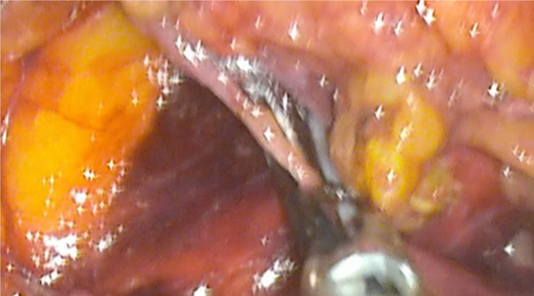
Figure 7 Identification of ureters with and without yellow enhancement[13].
A: With yellow enhancement; B: Without yellow enhancement. Citation: Suherman RC, Singh H, Aw DKL, Chong CXZ, Ng JL, Sivarajah S, Tan WJ, Foo FJ, Ladlad J, Khoo N, Tan CHM, Koh FH. The role of yellow enhancement in laparoscopy. Br J Surg 2024; 111. Copyright© The Authors 2024. Published by Oxford University Press on behalf of BJS Foundation Ltd. The authors have obtained the permission (Supplementary material).
- Citation: Singh H, Koh FHX. Golden vision: The potential of yellow enhancement in laparoscopic abdominal surgeries and surgical education. World J Gastrointest Endosc 2025; 17(7): 107872
- URL: https://www.wjgnet.com/1948-5190/full/v17/i7/107872.htm
- DOI: https://dx.doi.org/10.4253/wjge.v17.i7.107872


