©The Author(s) 2025.
World J Gastrointest Endosc. Jul 16, 2025; 17(7): 107645
Published online Jul 16, 2025. doi: 10.4253/wjge.v17.i7.107645
Published online Jul 16, 2025. doi: 10.4253/wjge.v17.i7.107645
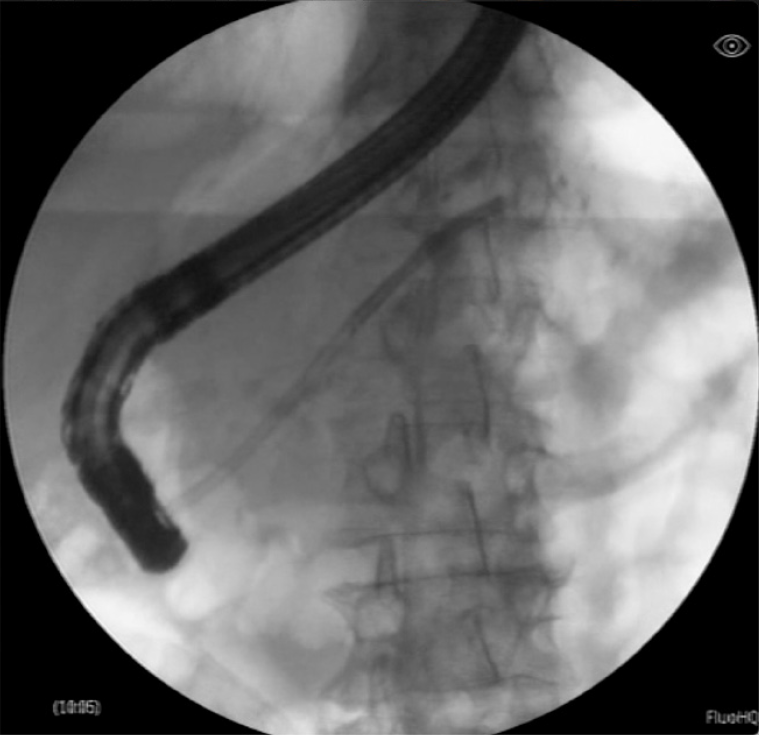
Figure 1
Fluoroscopic pancreatogram obtained during pancreatoscopy.
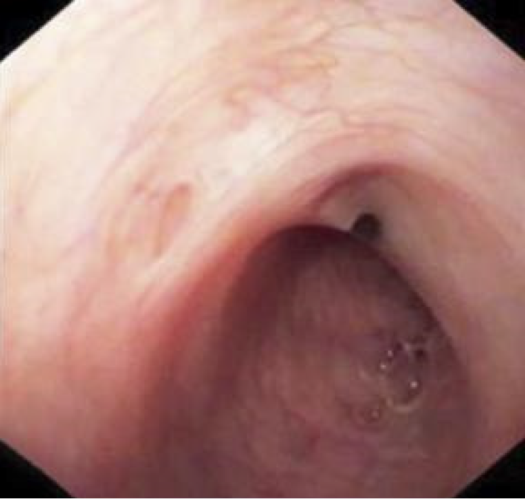
Figure 2
Normal pancreatic duct.
Figure 3 Intraductal papillary mucinous neoplasm.
A: Villous projections from a main-duct intraductal papillary mucinous neoplasm; B: SpyBite forceps used to perform a direct biopsy of a suspected intraductal papillary mucinous neoplasm; C: Intraductal papillary mucinous neoplasm and pancreatic stones.
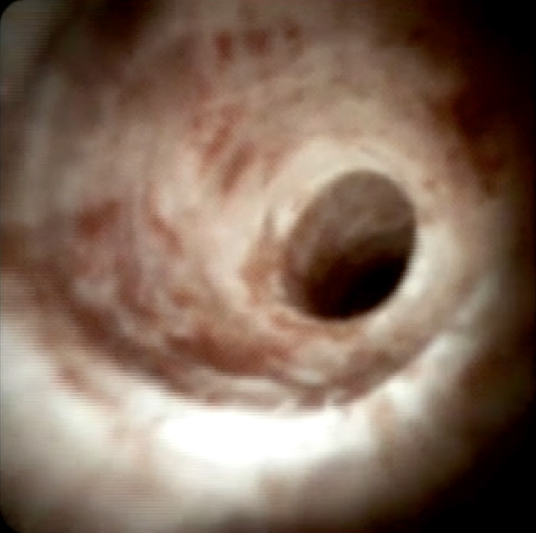
Figure 4
Main pancreatic duct stricture due to chronic pancreatitis.
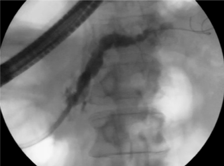
Figure 5
Fluoroscopy showing a tortuous pancreatic duct in chronic pancreatitis.
Figure 6 Stone lithotripsy.
A: Laser lithotripsy of a main pancreatic duct stone; B: Pancreatic side-branch stones; C: Electrohydraulic lithotripsy for main pancreatic duct stone fragmentation.
- Citation: Mansilla-Vivar R, Segovia-Vergara E, Pons-Beltrán V. Pancreatoscopy in the evaluation and management of pancreatic disorders. World J Gastrointest Endosc 2025; 17(7): 107645
- URL: https://www.wjgnet.com/1948-5190/full/v17/i7/107645.htm
- DOI: https://dx.doi.org/10.4253/wjge.v17.i7.107645


