©The Author(s) 2025.
World J Gastrointest Endosc. Jul 16, 2025; 17(7): 107587
Published online Jul 16, 2025. doi: 10.4253/wjge.v17.i7.107587
Published online Jul 16, 2025. doi: 10.4253/wjge.v17.i7.107587
Figure 1 Strasberg classification[15].
A: Bile leak from cystic duct stump or minor biliary radical in gallbladder fossa; B: Occluded right posterior sectoral duct; C: Bile leak from divided right posterior sectoral duct; D: Bile leak from main bile duct without major tissue loss; E1: Transected main bile duct with a stricture more than 2 cm from the hilus; E2: Transected main bile duct with a stricture less than 2 cm from the hilus; E3: Stricture of the hilus with right and left ducts in communication; E4: Stricture of the hilus with separation of right and left ducts; E5: Stricture of the main bile duct and the right posterior sectoral duct. Citation: Chun K. Recent classifications of the common bile duct injury. Korean J Hepatobiliary Pancreat Surg 2014; 18: 69-72. Copyright© The author(s) 2014. Published by The Korean Association of Hepato-Biliary-Pancreatic Surgery (Supplementary material).
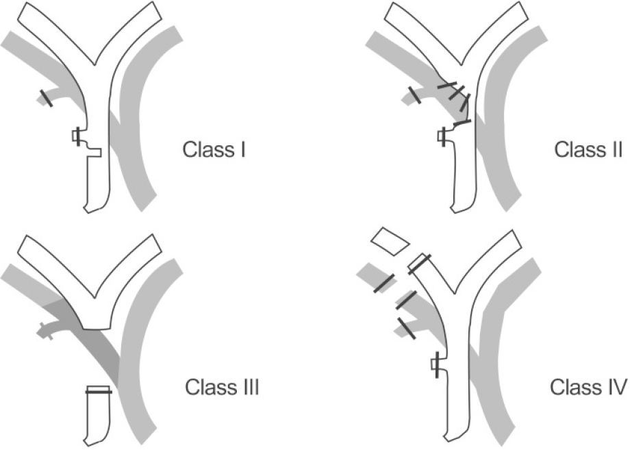
Figure 2 Stewart-Way classification[15].
Citation: Chun K. Recent classifications of the common bile duct injury. Korean J Hepatobiliary Pancreat Surg 2014; 18: 69-72. Copyright© The author(s) 2014. Published by The Korean Association of Hepato-Biliary-Pancreatic Surgery (Supplementary material).
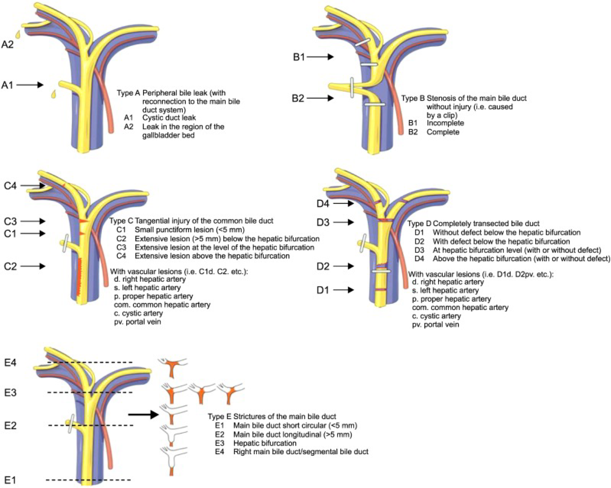
Figure 3 Hannover classification[15].
Citation: Chun K. Recent classifications of the common bile duct injury. Korean J Hepatobiliary Pancreat Surg 2014; 18: 69-72. Copyright© The author(s) 2014. Published by The Korean Association of Hepato-Biliary-Pancreatic Surgery (Supplementary material).
- Citation: Tringali A, Costa D, Ramai D. Endoscopic management of biliary leaks: Where are we now? World J Gastrointest Endosc 2025; 17(7): 107587
- URL: https://www.wjgnet.com/1948-5190/full/v17/i7/107587.htm
- DOI: https://dx.doi.org/10.4253/wjge.v17.i7.107587


