©The Author(s) 2025.
World J Gastrointest Endosc. Jul 16, 2025; 17(7): 107059
Published online Jul 16, 2025. doi: 10.4253/wjge.v17.i7.107059
Published online Jul 16, 2025. doi: 10.4253/wjge.v17.i7.107059
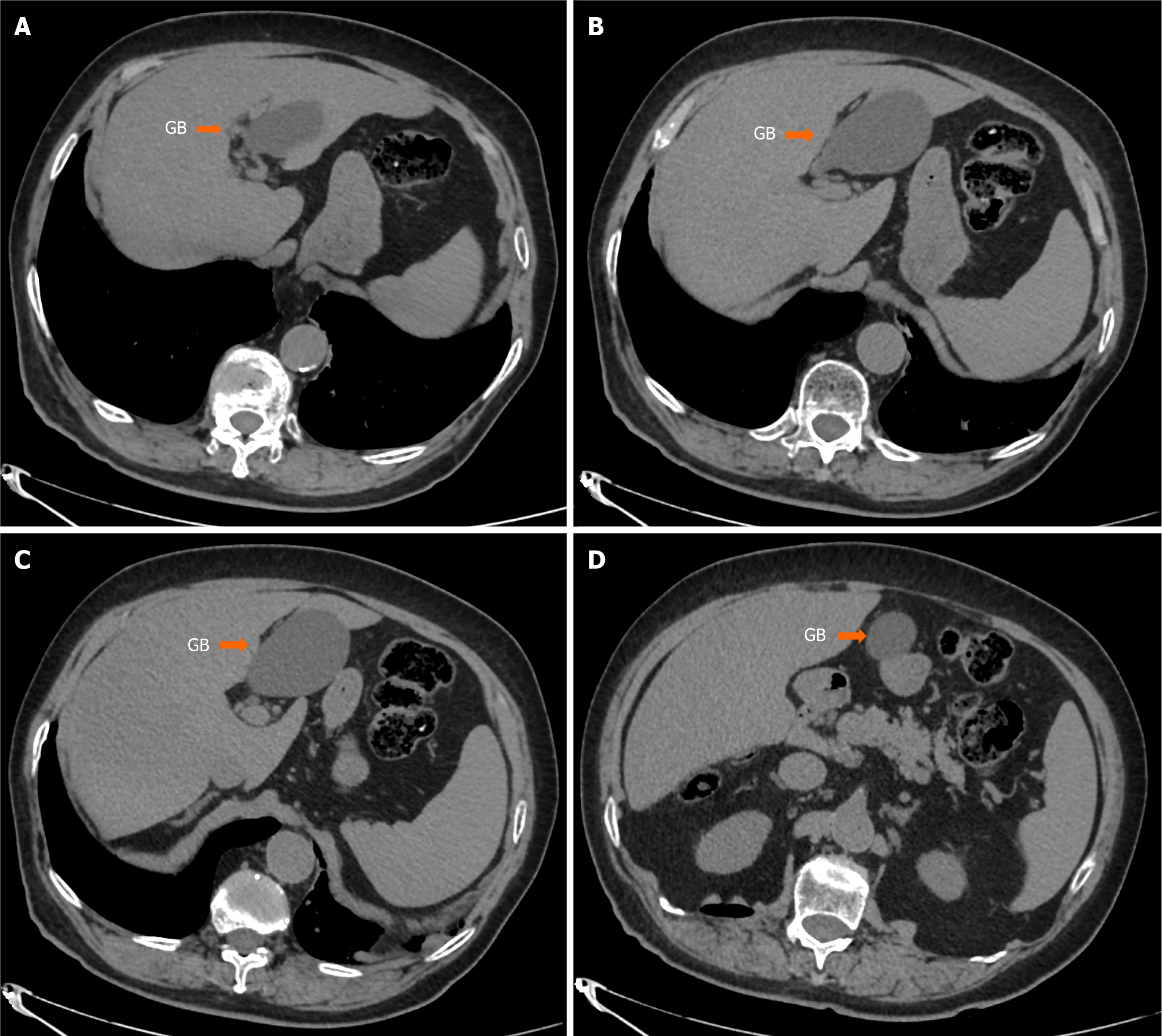
Figure 1 Abdominal computed tomography revealed suspected ectopic gallbladder.
A-D: Abdominal computed tomography revealed the gallbladder located below the round ligament of the liver, with a suspected attachment to the left lobe of the liver.
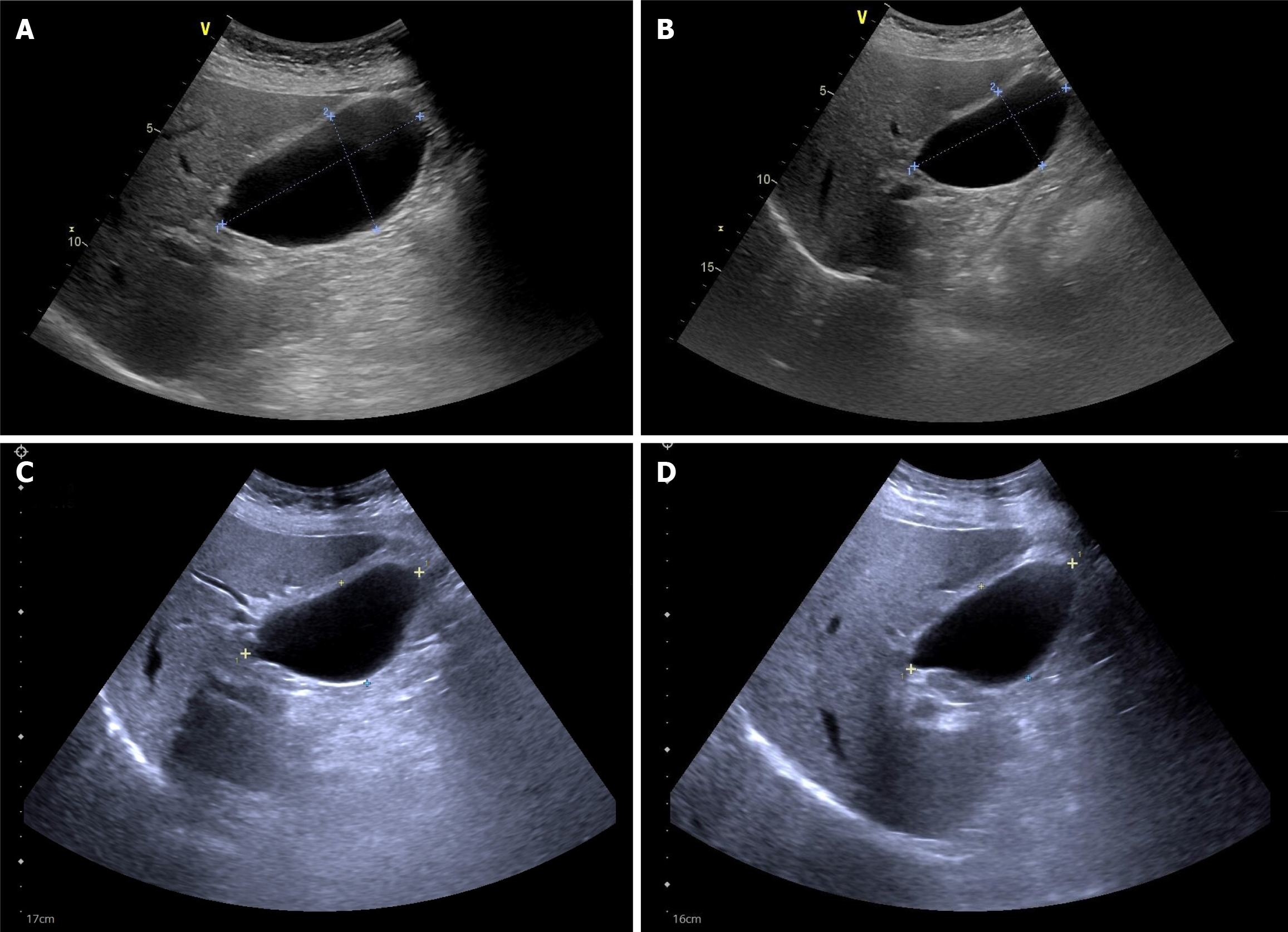
Figure 2 Fatty meal-stimulated, ultrasound-guided gallbladder emptying test revealed impaired gallbladder contraction.
A: Gallbladder was measured 8.1 cm × 4.8 cm × 4.6 cm before fatty meal; B: After 60 minutes, the gallbladder was 8.3 cm × 5.2 cm × 4.5 cm; C: To confirm the accuracy the test was repeated in the second day. At that time the gallbladder measured 8.0 cm × 4.4 cm × 4.3 cm before the fatty meal; D: 90 minutes after the meal, the gallbladder was 7.9 cm × 4.2 cm × 4.4 cm.
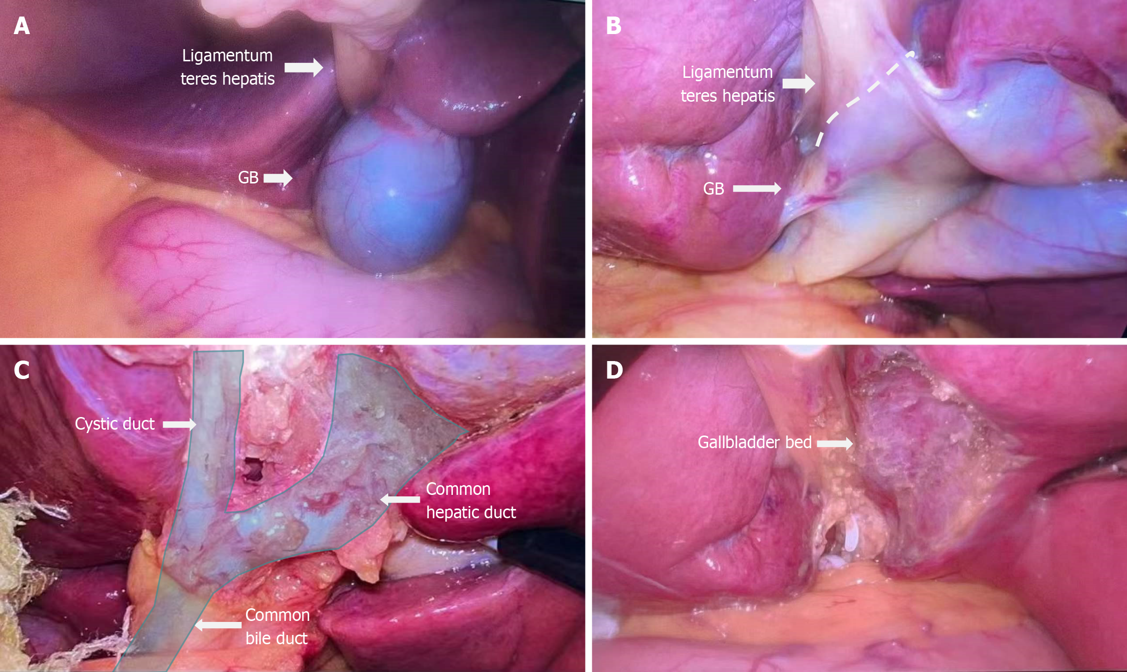
Figure 3 Left-sided gallbladder was confirmed during the operation.
A: After performing laparoscopic exploration in the abdomen, we discovered that the gallbladder was located below the ligamentum teres hepatis and left lateral lobe of the liver; B: We performed decompression of the gallbladder in order to visualize the boundaries of the gallbladder clearly. As a result, we observed that the majority of the gallbladder was attached to the left lateral lobe of the liver, with a smaller portion attached to the ligamentum teres.(The dashed line in the figure is the dividing line between the gallbladder and the round ligament of the liver); C: After dissecting the triangle of Calot, we found no abnormalities in the cystic duct, common hepatic duct, or common bile duct; D: Following gallbladder removal, we confirmed that the gallbladder bed was located on the left lateral lobe of the liver.
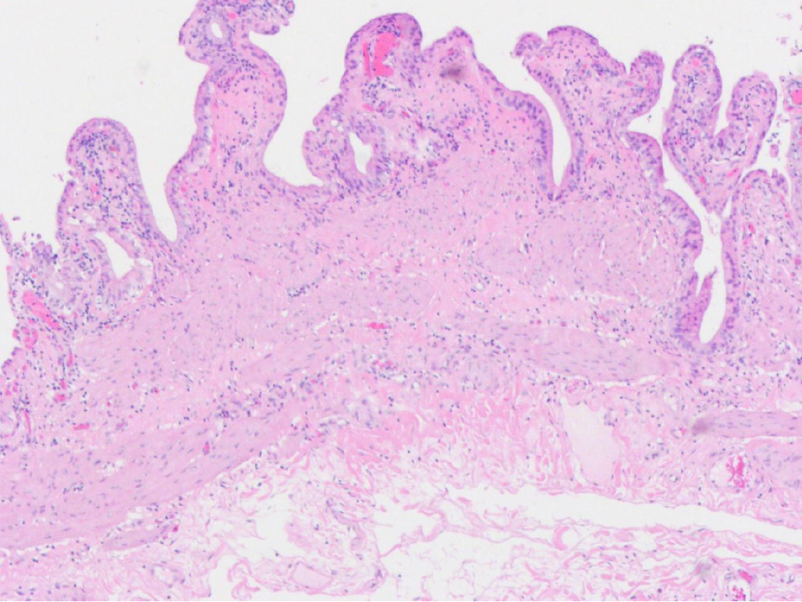
Figure 4 Pathology showed the thickness of the gallbladder wall was 0.
1-0.2 cm. The gallbladder was grayish-white, the mucosal surface was dark green, the mucosal surface was smooth, and the folds were visible. No significant abnormal pathological findings were found (hematoxylin-eosin staining, × 100).
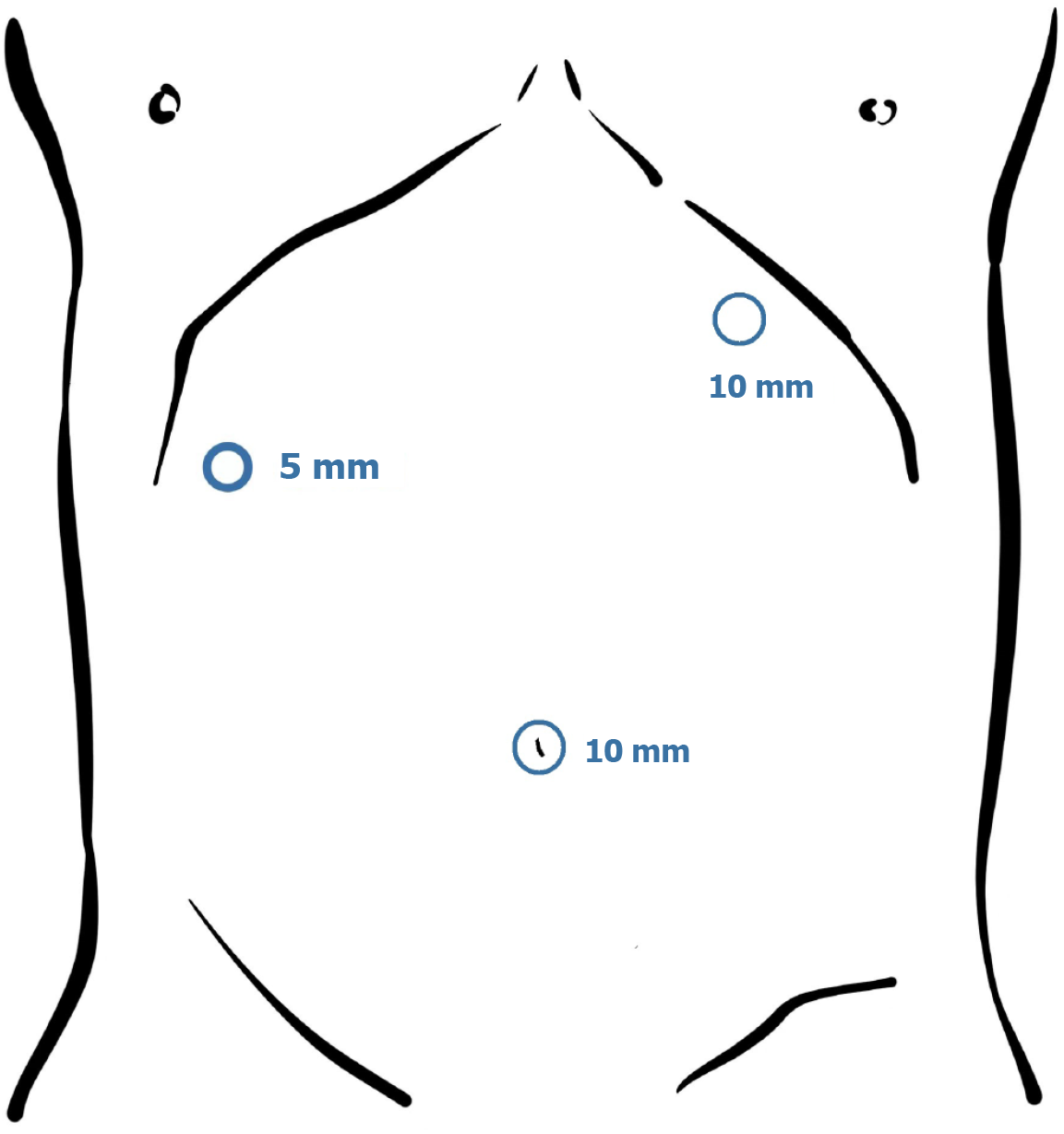
Figure 5 During surgery, a 10 mm trocar was initially inserted at the navel for observation.
Upon observing the left-sided gallbladder, a second 10 mm trocar was inserted at the midline of the left clavicle, 3 cm below the left costal margin. A 5 mm trocar was inserted at the right anterior axillary line, 3 cm below the right costal margin.
- Citation: Wu JR, Wang CC, Li BY, Li JH, Zhang T, Li ZY. Concomitant functional gallbladder disorder and left-sided gallbladder: A case report. World J Gastrointest Endosc 2025; 17(7): 107059
- URL: https://www.wjgnet.com/1948-5190/full/v17/i7/107059.htm
- DOI: https://dx.doi.org/10.4253/wjge.v17.i7.107059


