©The Author(s) 2025.
World J Gastrointest Endosc. Jul 16, 2025; 17(7): 105773
Published online Jul 16, 2025. doi: 10.4253/wjge.v17.i7.105773
Published online Jul 16, 2025. doi: 10.4253/wjge.v17.i7.105773
Figure 1 A stone at Kasai portoenterostomy sites in computed tomography and magnetic resonance imaging.
A: The axial images of the abdominal computed tomography scan, taken in the local hospital before transfer, show a hyperdense lesion (arrows) at the hepatic hilum suggesting bile sludge or stone; B: The coronal image of the abdominal magnetic resonance imaging taken at our hospital after transfer shows a T2 low-signal intensity lesion (arrow) at the Kasai portoenterostomy site.
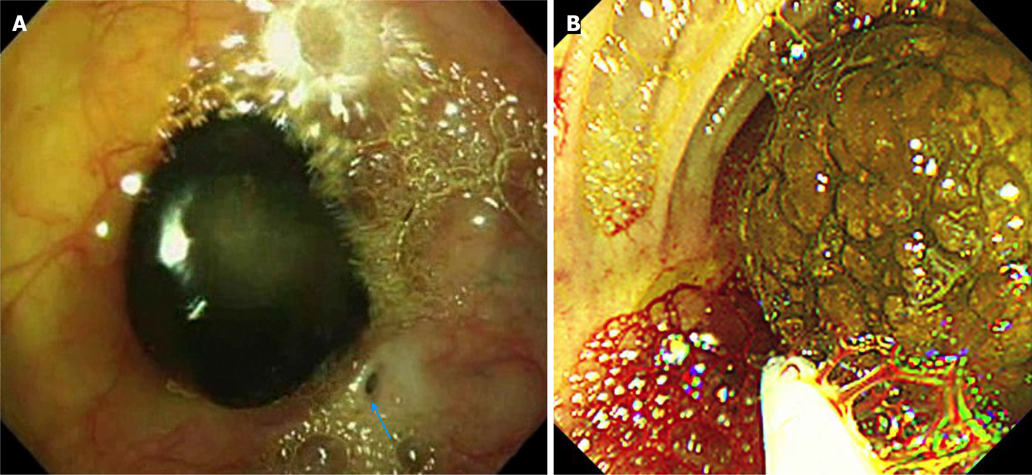
Figure 2 Intraoperative endoscopic view of stone.
A: Intraoperative endoscopic findings in an unpublished previous case of a Kasai portoenterostomy (KPE) site stone, experienced 14 years ago, showing a black pigmented stone hanging like a stalactite from the KPE site and a tiny opening of a bile ductule (arrow). Endoscopic removal of the stone resolved the cholangitis and prevented further development of cholangitis in this case; B: Intraoperative endoscopic findings in the current case showing a large thick bile sludge-like lump hanging from the KPE site, which was easily broken during endoscopic removal. Three minutes after starting the endoscope examination, a sudden fatal air embolism occurred, and the patient died.
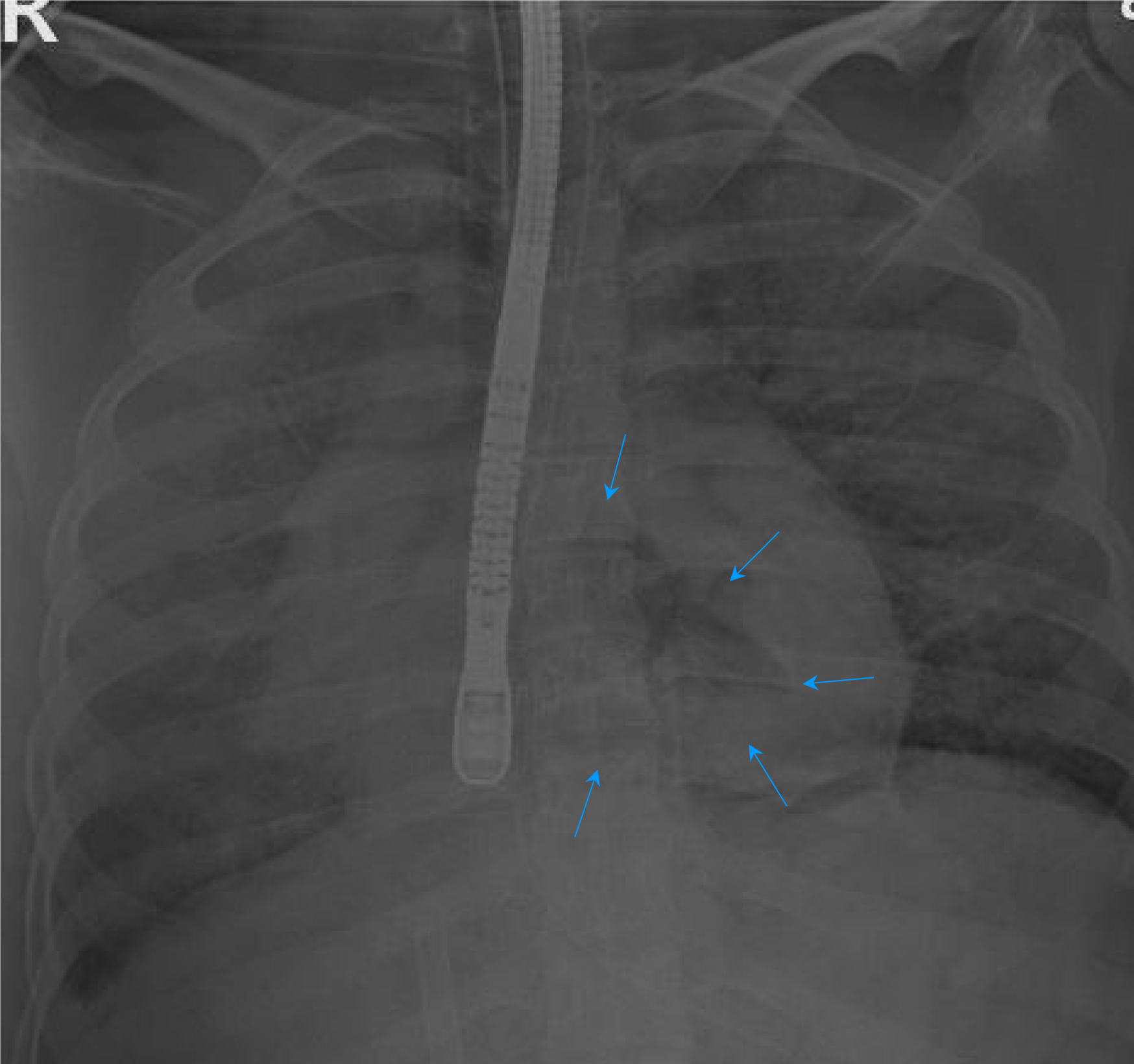
Figure 3
Air embolism in cardiac chamber (arrow) confirmed with chest X-ray taken at the operating room.
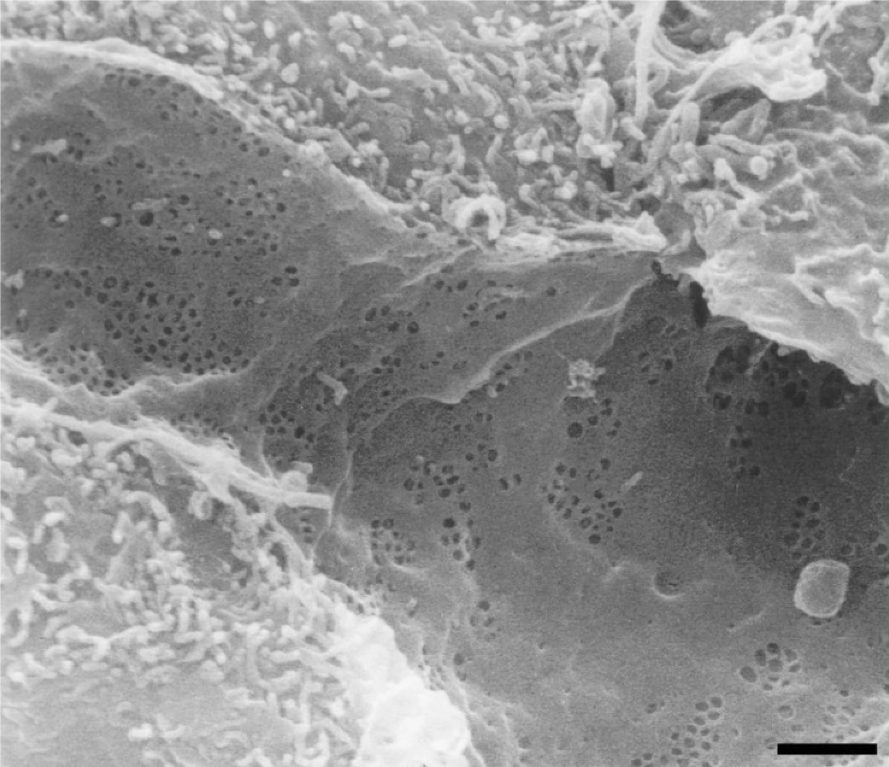
Figure 4 Low magnification scanning electron micrograph of the sinusoidal endothelium from a rat liver showing the fenestrated wall.
Shown is the clustering of fenestrae in sieve plates. Scale bar: 1 μm. Citation: Braet F, Wisse E. Structural and functional aspects of liver sinusoidal endothelial cell fenestrae: a review. Comp Hepatol 2002; 1(1): 1. Copyright ©The Author(s) 2019. Published by BioMed Central Ltd[7].
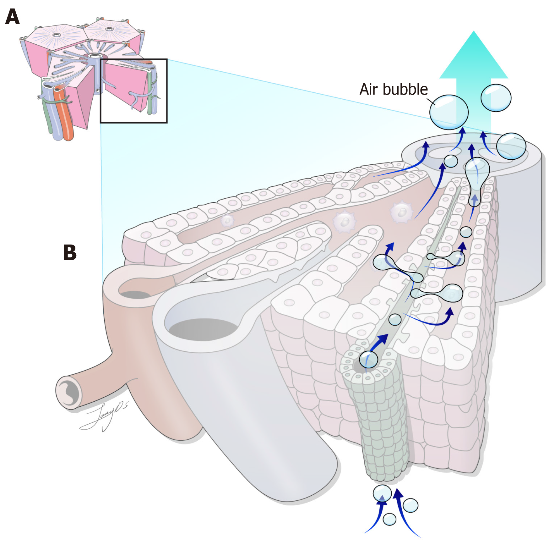
Figure 5
When high-pressure air is insufflated into a closed blind loop during intestinal endoscopy, as in patients with Kasai portoenterostomy, air may enter the bile canaliculi and pass into the bloodstream through these tiny pores of liver sinusoid endothelial cells, potentially leading to fatal air embolism.
- Citation: Shin SY, Yeon HJ, Lee SO, Lee JR, Leem G, Han SJ. Fatal air embolism during intestinal endoscopy in Kasai portoenterostomy for biliary atresia: A case report. World J Gastrointest Endosc 2025; 17(7): 105773
- URL: https://www.wjgnet.com/1948-5190/full/v17/i7/105773.htm
- DOI: https://dx.doi.org/10.4253/wjge.v17.i7.105773


