©The Author(s) 2024.
World J Gastrointest Endosc. Dec 16, 2024; 16(12): 678-685
Published online Dec 16, 2024. doi: 10.4253/wjge.v16.i12.678
Published online Dec 16, 2024. doi: 10.4253/wjge.v16.i12.678
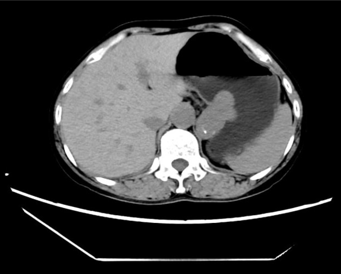
Figure 1 Computed tomography revealed an irregular submucosal mass in the gastric fundus and cardia, some of which protruded out of the cavity, with a size of about 4.
5 cm × 3.2 cm × 4.9 cm and a clear boundary. Nodular calcification was observed within the lesion, with a computed tomography (CT) value of about 42 HU, and mild enhancement was observed. The tertiary CT values were about 52 HU, 54 HU, and 55 HU, and linear enhancement was observed at the lateral edge of the lesion. There were no obvious enlarged lymph nodes around the lesion.
Figure 2 Endoscopic appearance and resection process of the lesion.
A: Under the gastroscope, a submucosal mass was found in the fundus of the stomach, with a maximum length of about 5 cm, and the surface was depressed and locally rough, involving the cardia; B: The lesion along the submucosa and muscle layer was peeled to expose the tumor; C: Dissection of the lesion was completed; D: The longest wound after lesion dissection was about 8 cm; E: The wound was closed using a double pouches suture technique; F: The size of the lesion was about 8 cm × 5 cm, and after being restored to its original shape, it was removed in segments.
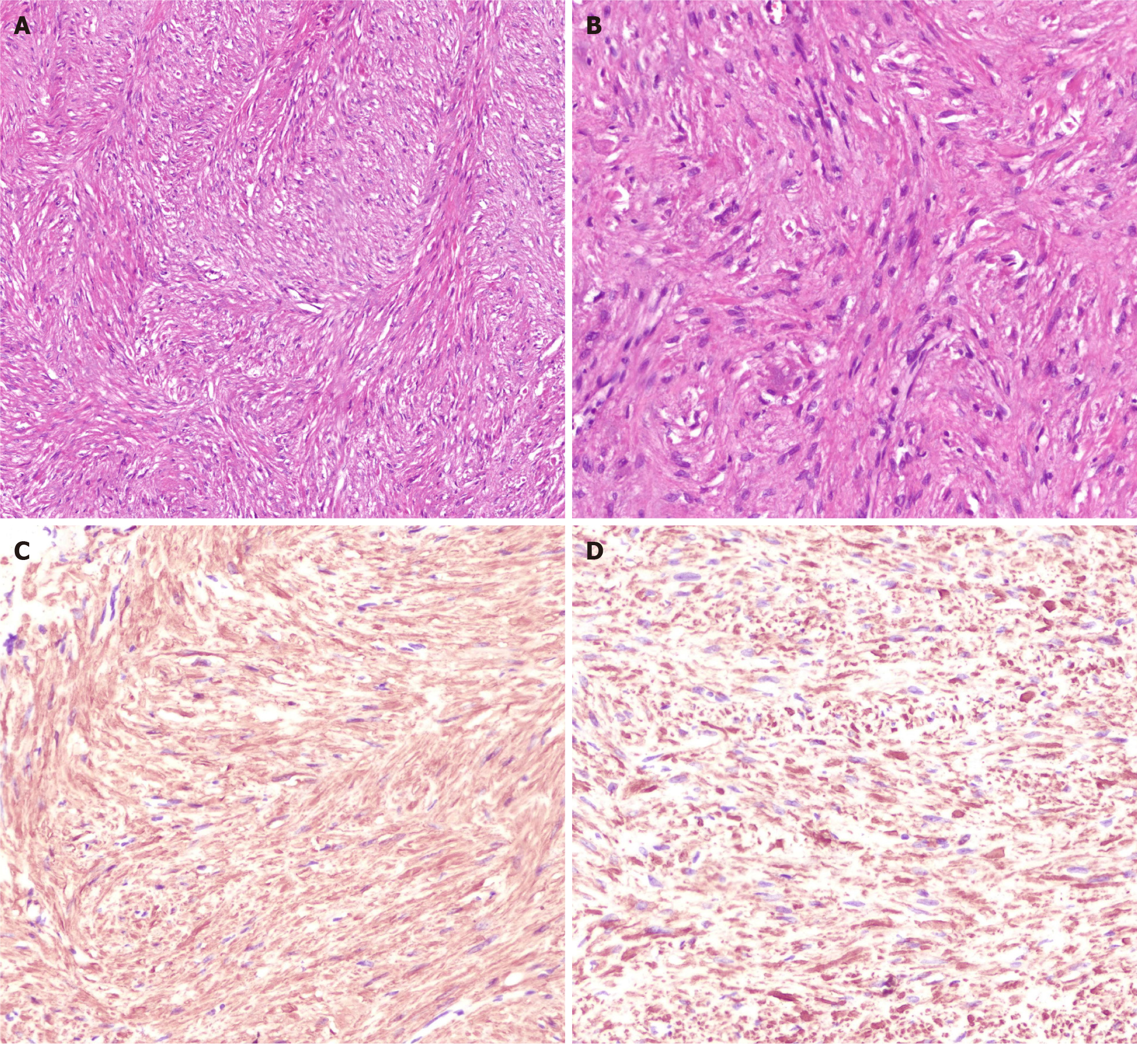
Figure 3 Histopathology and immunohistochemistry suggested leiomyoma.
A: HE staining showed that the tumor cells were long spindle-shaped, and the cytoplasm was rich in red stain and was arranged in bundles or interleaved shapes (HE × 20); B: At high magnification, HE staining showed that the nucleus was long rod-shaped (longitudinal cut) or round oval (transverse cut), located in the center of the cytoplasm, and the nucleolus was not obvious. The nuclear morphology was mild and heterotypic, the chromatin was uniform, and the mitosis was rare (HE × 40); C: Immunohistochemistry showed positive SMA (× 40); D: Immunohistochemistry indicated positive Desmin (× 40).
- Citation: Li P, Tang GM, Li PL, Zhang C, Wang WQ. Endoscopic resection of a giant irregular leiomyoma in fundus and cardia: A case report. World J Gastrointest Endosc 2024; 16(12): 678-685
- URL: https://www.wjgnet.com/1948-5190/full/v16/i12/678.htm
- DOI: https://dx.doi.org/10.4253/wjge.v16.i12.678


