Copyright
©The Author(s) 2023.
World J Gastrointest Endosc. Apr 16, 2023; 15(4): 273-284
Published online Apr 16, 2023. doi: 10.4253/wjge.v15.i4.273
Published online Apr 16, 2023. doi: 10.4253/wjge.v15.i4.273
Figure 1 Endoscopic ultrasound characteristic of solid pseudopapillary tumors.
Figure 2 Endoscopic ultrasound.
A: A large heterogeneous solid pseudopapillary neoplasm in the pancreatic head; B: A large heterogeneous solid pseudopapillary neoplasm with calcific spots in the pancreatic head; C: A cystic solid pseudopapillary neoplasm in the pancreatic body.
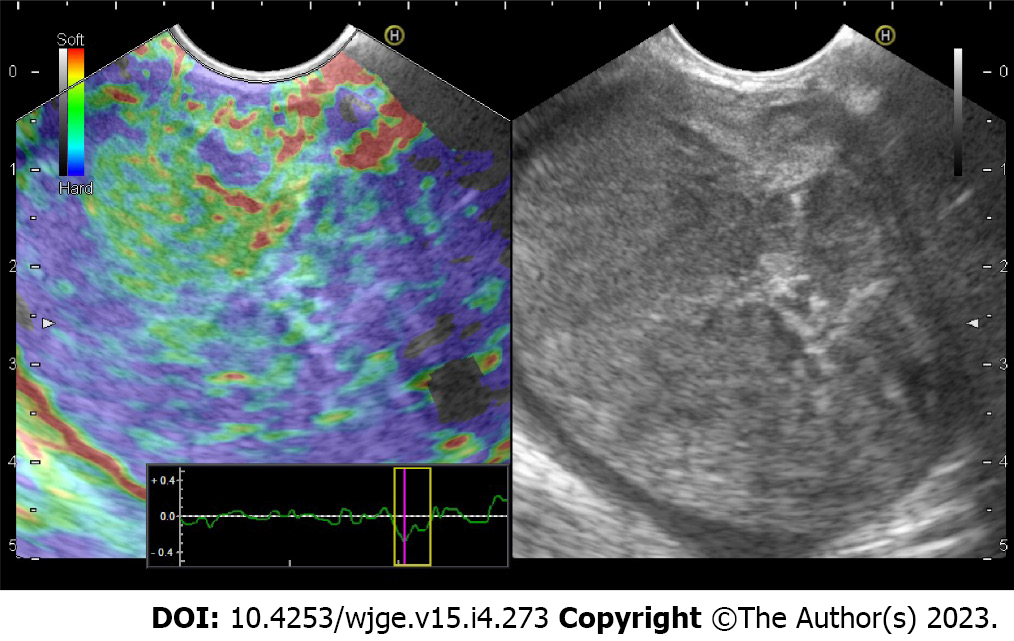
Figure 3 A large heterogeneous firm solid pseudopapillary neoplasm in the pancreatic head with dominant blue color denoting grade 3 Elasticity score.
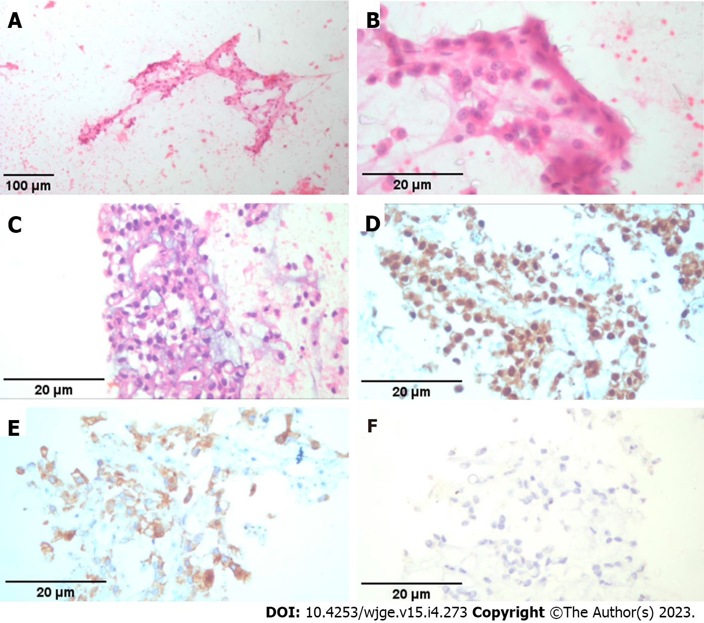
Figure 4 Smears of solid pseudopapillary pancreatic tumor.
A and B: Clusters of uniform epithelioid cells arranged in a vague papillary like formation (Hematoxylin & Eosin × 40); C: Cell block of same tumor (Hematoxylin & Eosin × 400); D: Positive nuclear B-Catenin immunoreaction in tumor cells (Hematoxylin & Eosin × 400); E: Positive cytoplasmic Synatophysin immunoreaction in tumor cells (Hematoxylin & Eosin × 400); F: Negative Chromogranin immunoreaction (Hematoxylin & Eosin × 400).
Figure 5 A post-operative specimen of mixed solid pseudopapillary neoplasm with solid and cystic areas.
- Citation: Pawlak KM, Tehami N, Maher B, Asif S, Rawal KK, Balaban DV, Tag-Adeen M, Ghalim F, Abbas WA, Ghoneem E, Ragab K, El-Ansary M, Kadir S, Amin S, Siau K, Wiechowska-Kozlowska A, Mönkemüller K, Abdelfatah D, Abdellatef A, Lakhtakia S, Okasha HH. Role of endoscopic ultrasound in the characterization of solid pseudopapillary neoplasm of the pancreas. World J Gastrointest Endosc 2023; 15(4): 273-284
- URL: https://www.wjgnet.com/1948-5190/full/v15/i4/273.htm
- DOI: https://dx.doi.org/10.4253/wjge.v15.i4.273


