©The Author(s) 2023.
World J Gastrointest Endosc. Apr 16, 2023; 15(4): 265-272
Published online Apr 16, 2023. doi: 10.4253/wjge.v15.i4.265
Published online Apr 16, 2023. doi: 10.4253/wjge.v15.i4.265
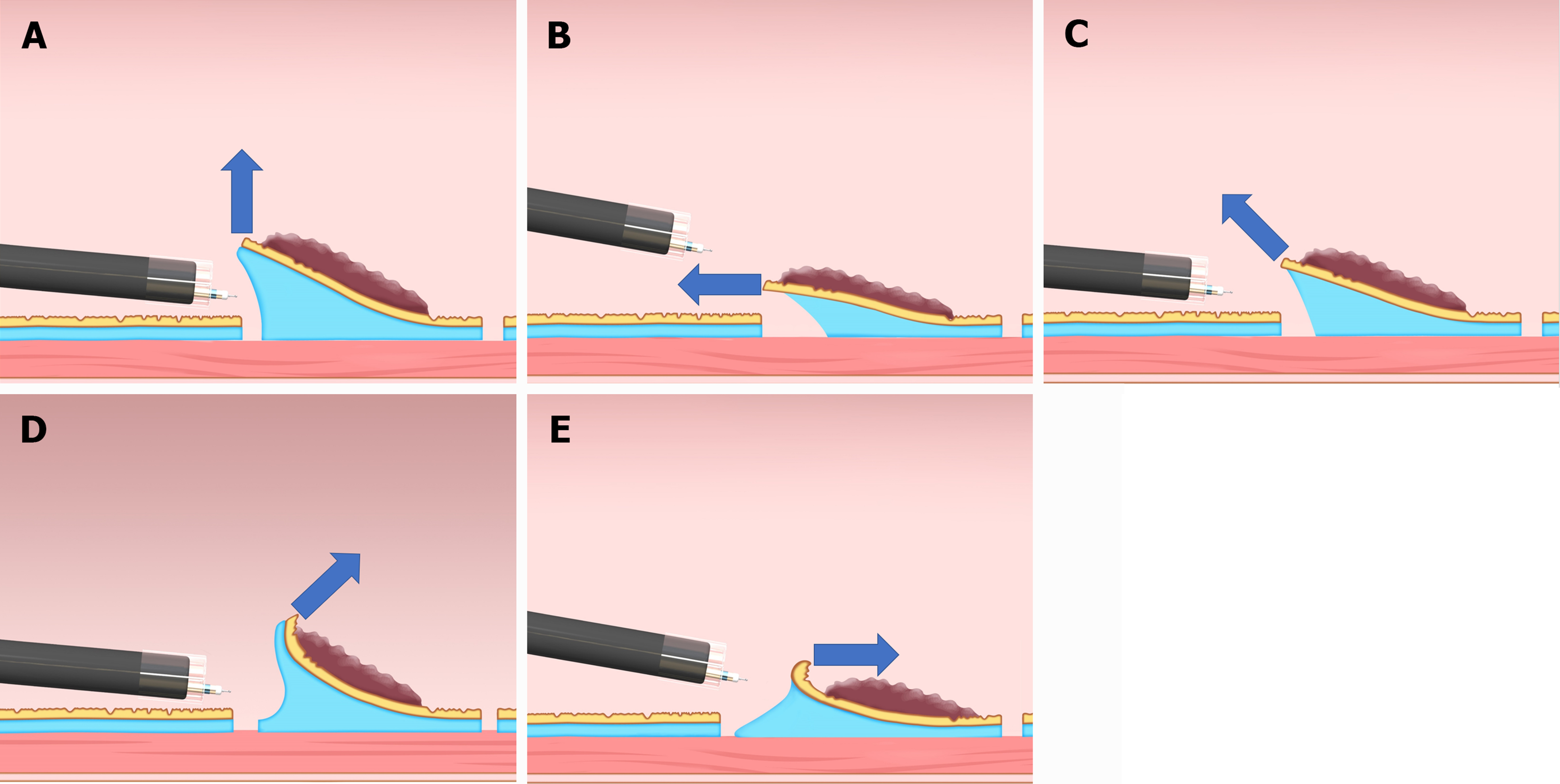
Figure 1 Classification of the traction direction.
A: Vertical traction; B: Proximal traction; C: Diagonally proximal traction; D: Diagonally distal traction; E: Distal traction. Citation: Reprinted from Mitsuru Nagata. Advances in traction methods for endoscopic submucosal dissection: What is the best traction method and traction direction? World Journal of Gastroenterology 2022; 28: 1-22. Copyright ©Mitsuru Nagata 2022. Published by Baishideng Publishing Group Inc[8].
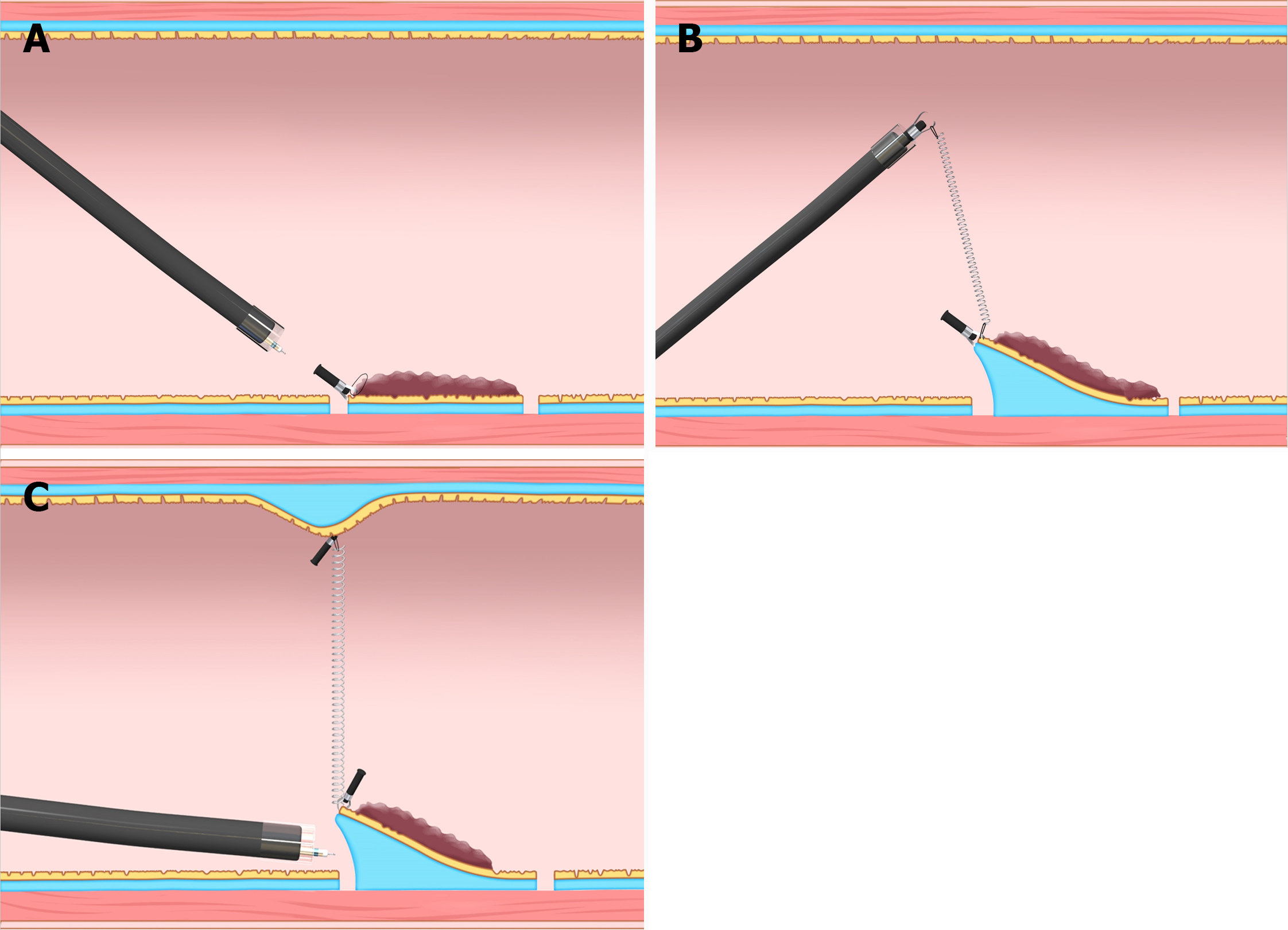
Figure 2 Internal traction method using the spring-and-loop with clip (Zeon Medical, Tokyo, Japan).
A: The spring-and-loop with clip (S–O clip) is attached to the lesion; B: The regular clip anchors the loop part of the S–O clip on the gastrointestinal wall; C: The extension of the spring provides traction on the lesion. The traction direction can be controlled by the anchor site. Citation: Figure 2 reprinted from Nagata M, Fujikawa T, Munakata H. Comparing a conventional and a spring-and-loop with clip traction method of endoscopic submucosal dissection for superficial gastric neoplasms: a randomized controlled trial (with videos). Gastrointestinal Endoscopy 2021; 93: 1097-1109. Copyright ©2021 American Society for Gastrointestinal Endoscopy. Published by Elsevier Inc[10].
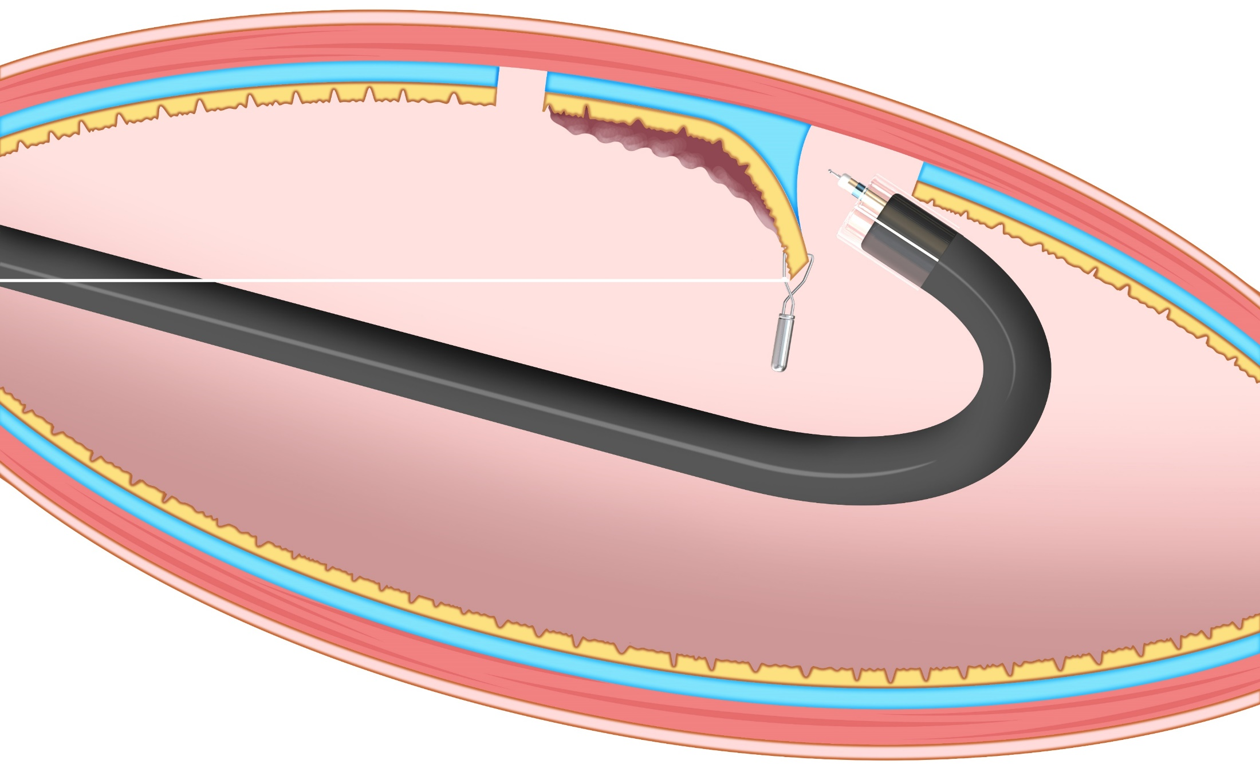
Figure 3 Clip-with-line method.
This method provides traction for the lesion by pulling the line. The traction direction is limited to the direction in which the line is pulled. Citation: Figure 3 reprinted from Mitsuru Nagata. Advances in traction methods for endoscopic submucosal dissection: What is the best traction method and traction direction? World Journal of Gastroenterology 2022; 28: 1–22. Copyright ©Mitsuru Nagata 2022. Published by Baishideng Publishing Group Inc[8].
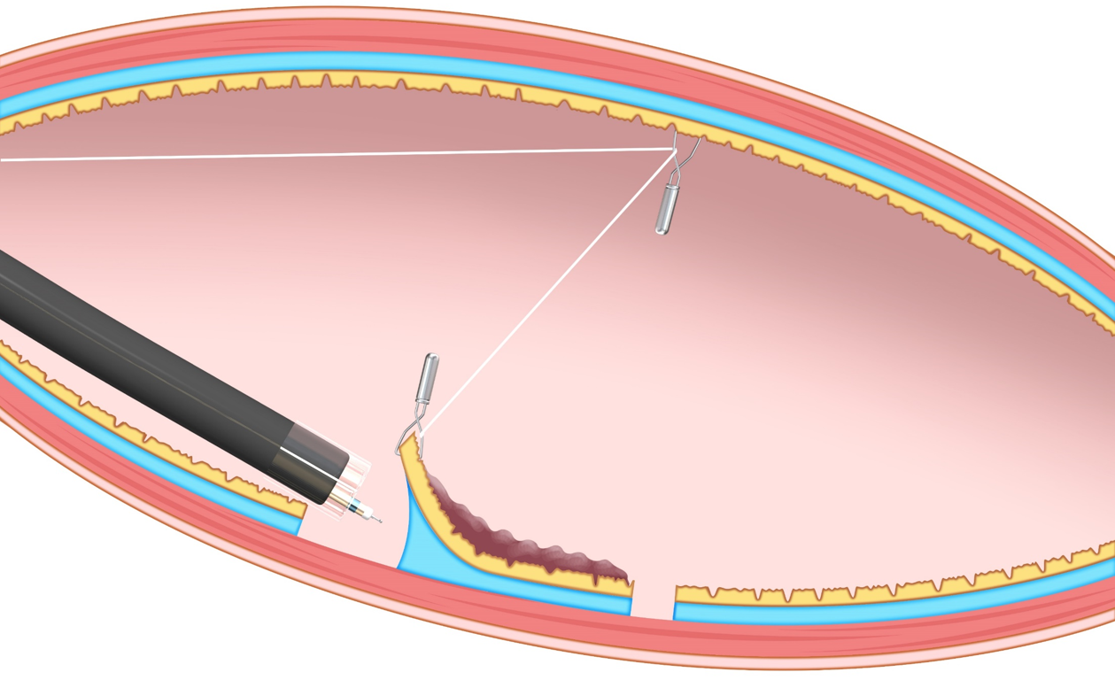
Figure 4 Pulley method.
This method is the modified clip-with-line method, which can provide traction in any direction depending on the pulley site. Citation: Reprinted from Mitsuru Nagata. Advances in traction methods for endoscopic submucosal dissection: What is the best traction method and traction direction? World Journal of Gastroenterology 2022; 28: 1–22. Copyright ©Mitsuru Nagata 2022. Published by Baishideng Publishing Group Inc[8].
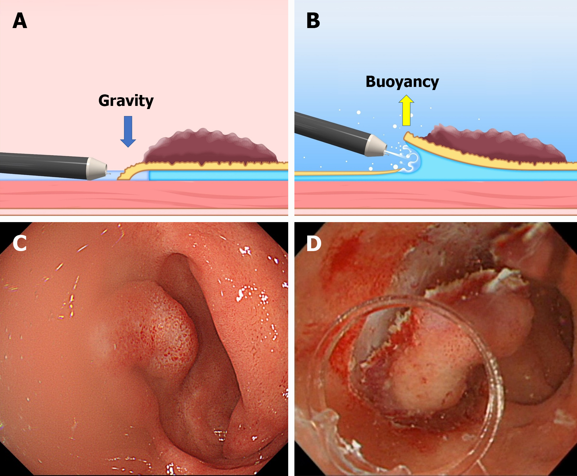
Figure 5 The difference between the conventional endoscopic submucosal dissection and the underwater endoscopic submucosal dissection.
A and C: The conventional endoscopic submucosal dissection for the lesion is located at the gravitational lower side. Gravity obstructs the opening of the mucosal flap. Incomplete submersion deteriorates the visual field; B and D: The underwater condition aids the opening of the mucosal flap by buoyancy. Water pressure from the endoscope (using its water supply function) also assists in opening the mucosal flap. Complete submersion improves the visual field. Citation: Figure 5A and B reprinted from Mitsuru Nagata. Advances in traction methods for endoscopic submucosal dissection: What is the best traction method and traction direction? World Journal of Gastroenterology 2022; 28: 1–22. Copyright ©Mitsuru Nagata 2022. Published by Baishideng Publishing Group Inc[8]. Figure 5C and D reprinted from Mitsuru Nagata. Underwater endoscopic submucosal dissection in saline solution using a bent-type knife for duodenal tumor. VideoGIE 2018; 3: 375–377. Copyright ©2018 American Society for Gastrointestinal Endoscopy. Published by Elsevier Inc[20].
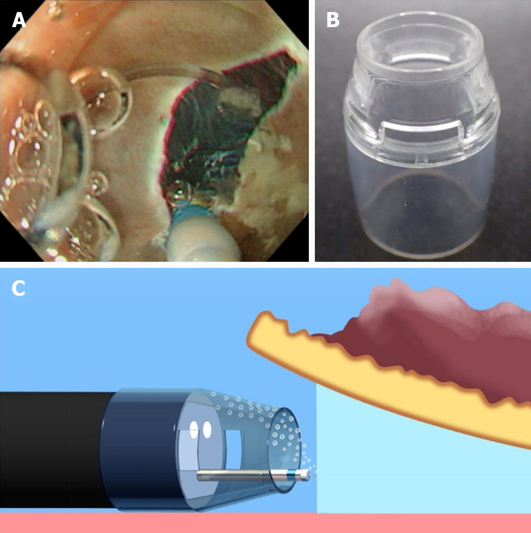
Figure 6 Underwater endoscopic submucosal dissection with a conventional tapered hood.
A: Accumulation of air bubbles in the hood causes visual field impairment during underwater endoscopic submucosal dissection; B: A conventional tapered hood (ST Hood Short Type; Fujifilm Medical, Tokyo, Japan). This hood has narrow holes in its sides to remove the liquid via the capillary phenomenon, which is inversely proportional to the cross-sectional area of the hole; C: When a conventional tapered hood is used, removal of air bubbles can sometimes be difficult due to the narrow hood tip opening and holes in its sides. Citation: Reprinted from Mitsuru Nagata. Tapered hood with wide holes in its sides for efficient air bubble removal during underwater endoscopic submucosal dissection. Digestive Endoscopy 2022; 34: 654. ©2022 Japan Gastroenterological Endoscopy Society. Published by John Wiley & Sons Australia, Ltd[21].
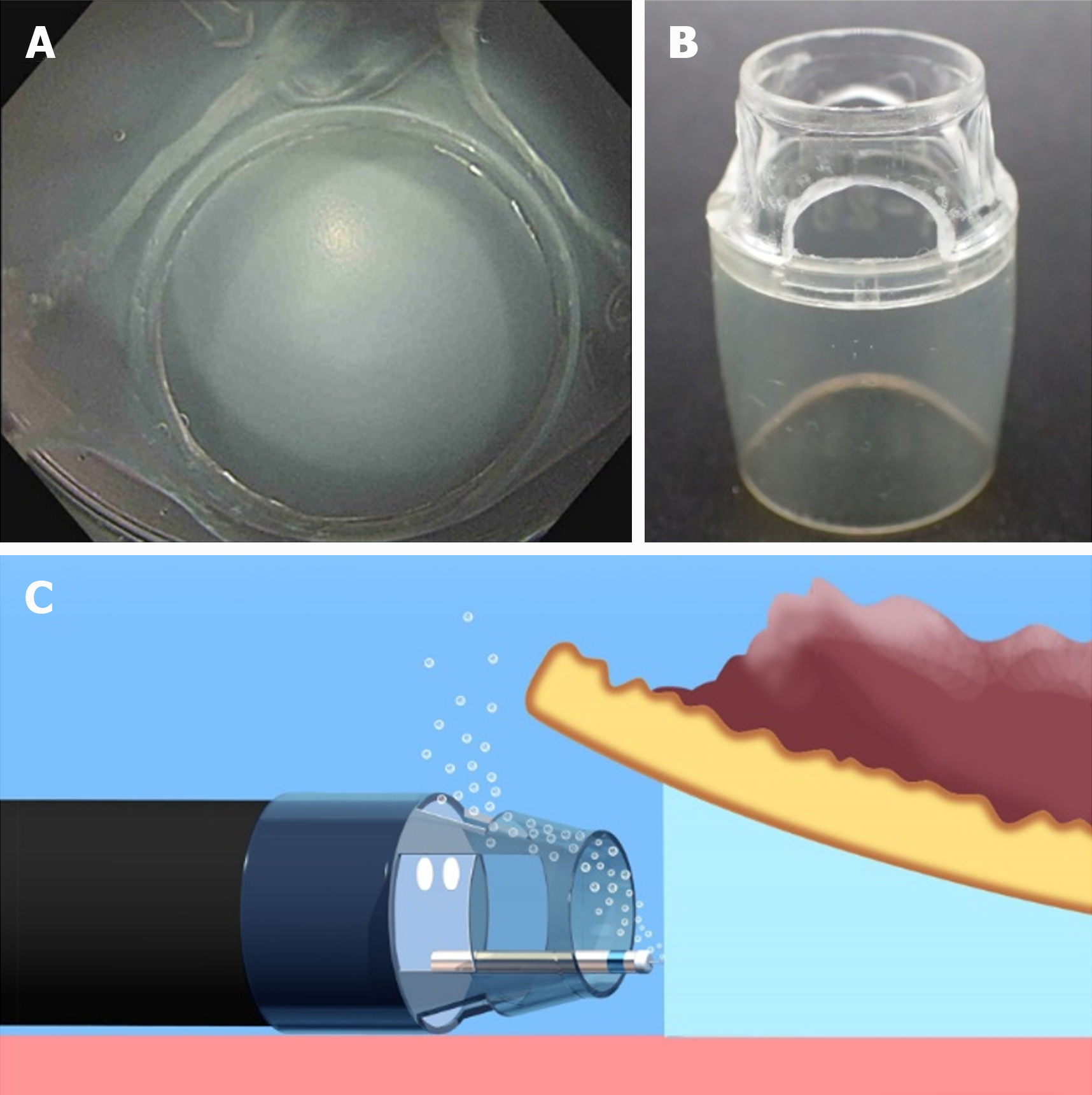
Figure 7 A tapered hood with wide holes in its sides for efficient air bubble removal during underwater endoscopic submucosal dissection.
A: An endoscopic view when a tapered tip hood with wide holes in its sides is attached; B: A tapered hood (ST Hood Short Type; Fujifilm Medical) with wide holes in its sides for air bubble removal during underwater endoscopic submucosal dissection. As the tip of an ST Hood Short Type is made of polycarbonate resin, wide holes in its sides can be made by hand using a commercially available router (RTD35ACL; TACKLIFE); C: Wide holes in the sides of the tapered hood can facilitate air bubble removal. Citation: Reprinted from Mitsuru Nagata. Tapered hood with wide holes in its sides for efficient air bubble removal during underwater endoscopic submucosal dissection. Digestive Endoscopy 2022; 34: 654. ©2022 Japan Gastroenterological Endoscopy Society. Published by John Wiley & Sons Australia, Ltd[21].
- Citation: Nagata M. Device-assisted traction methods in colorectal endoscopic submucosal dissection and options for difficult cases. World J Gastrointest Endosc 2023; 15(4): 265-272
- URL: https://www.wjgnet.com/1948-5190/full/v15/i4/265.htm
- DOI: https://dx.doi.org/10.4253/wjge.v15.i4.265


