©The Author(s) 2023.
World J Gastrointest Endosc. Dec 16, 2023; 15(12): 690-698
Published online Dec 16, 2023. doi: 10.4253/wjge.v15.i12.690
Published online Dec 16, 2023. doi: 10.4253/wjge.v15.i12.690
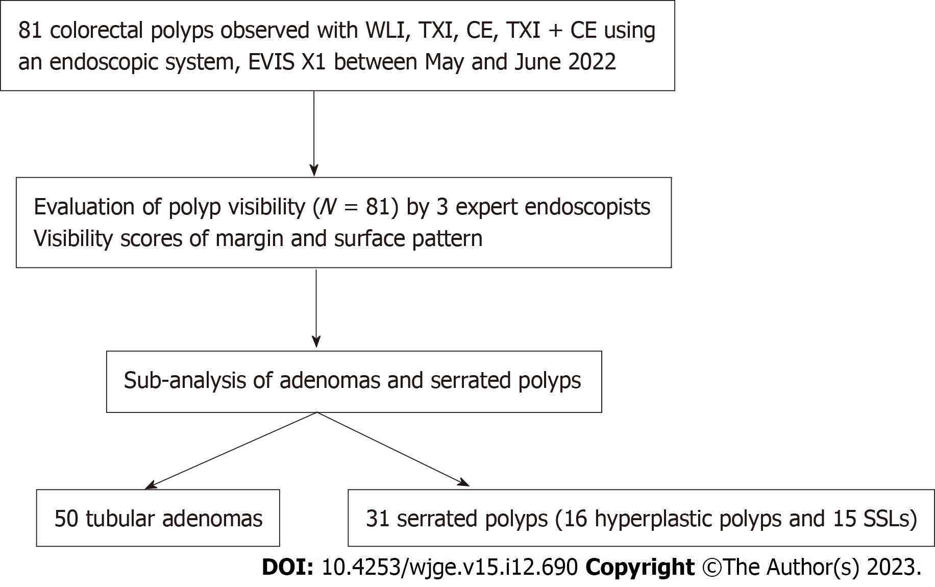
Figure 1 Flowchart for the study design.
WLI: White light imaging; TXI: Texture and color enhancement imaging; CE: Chromoendoscopy; SSL: Sessile serrated lesion.
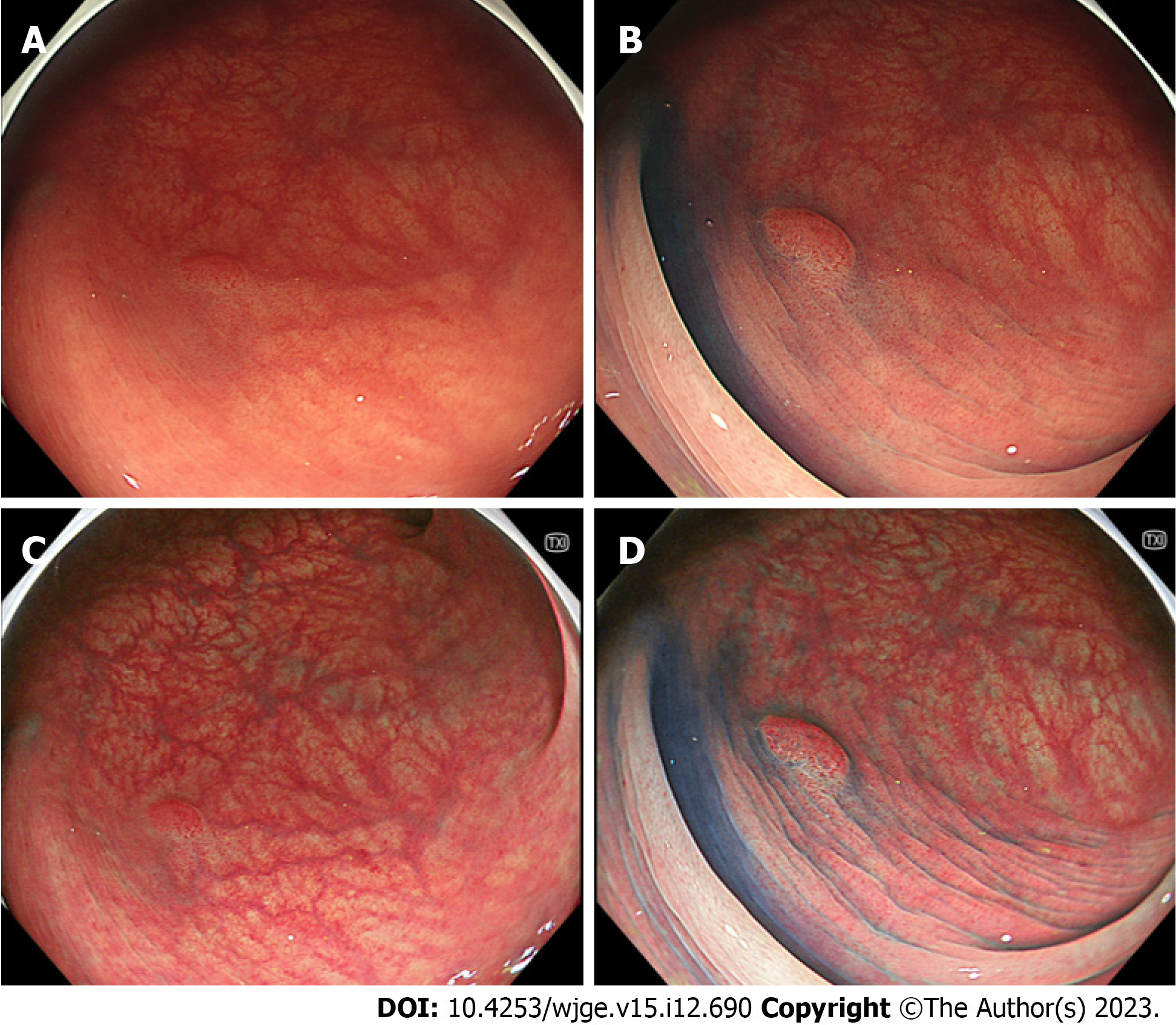
Figure 2 Representative images of adenoma.
A: White light imaging; B: Chromoendoscopy; C: Texture and color enhancement imaging; D: Chromoendoscopy and texture and color enhancement imaging.
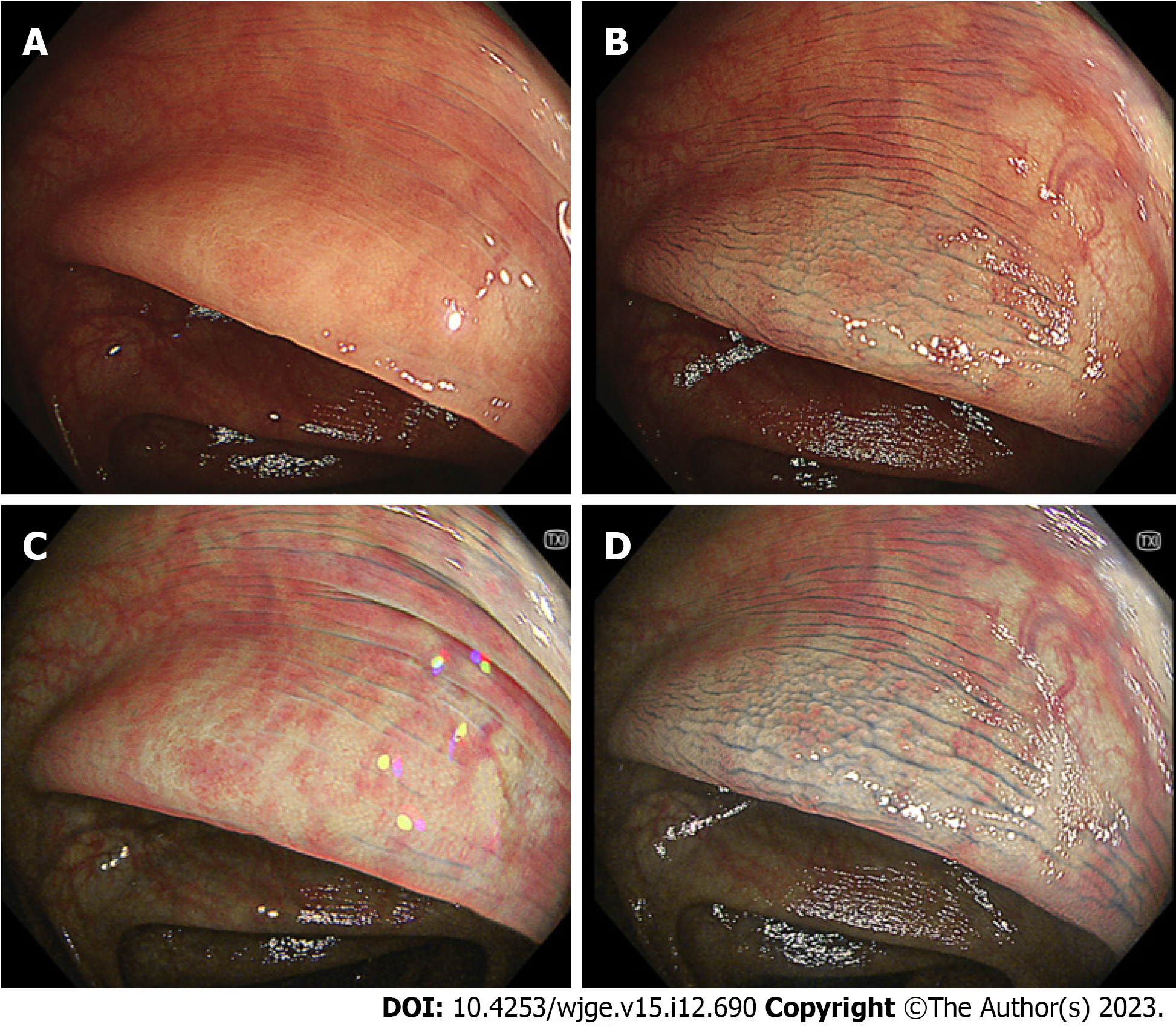
Figure 3 Representative images of serrated polyp (sessile serrated lesion).
A: White light imaging; B: Chromoendoscopy; C: Texture and color enhancement imaging; D: Chromoendoscopy and texture and color enhancement imaging.
- Citation: Hiramatsu T, Nishizawa T, Kataoka Y, Yoshida S, Matsuno T, Mizutani H, Nakagawa H, Ebinuma H, Fujishiro M, Toyoshima O. Improved visibility of colorectal tumor by texture and color enhancement imaging with indigo carmine. World J Gastrointest Endosc 2023; 15(12): 690-698
- URL: https://www.wjgnet.com/1948-5190/full/v15/i12/690.htm
- DOI: https://dx.doi.org/10.4253/wjge.v15.i12.690


