©The Author(s) 2023.
World J Gastrointest Endosc. Nov 16, 2023; 15(11): 634-640
Published online Nov 16, 2023. doi: 10.4253/wjge.v15.i11.634
Published online Nov 16, 2023. doi: 10.4253/wjge.v15.i11.634
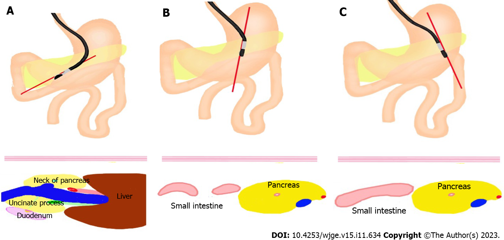
Figure 1 Endoscopic ultrasound scans the suitable bowel to do endoscopic ultrasound- guided gastroenterostomy.
A: We scan the confluence of splenic vein and superior mesenteric vein, we can see the neck of pancreas, uncinate process and the second part of duodenum behind the uncinate process; B: We slightly rotate the endoscope, then we can see the short-axis view of bowel near to stomach and below the pancreas; C: When we continue to rotate the endoscope, we can see the long-axis view of bowel.
- Citation: Wang J, Hu JL, Sun SY. Endoscopic ultrasound guided gastroenterostomy: Technical details updates, clinical outcomes, and adverse events. World J Gastrointest Endosc 2023; 15(11): 634-640
- URL: https://www.wjgnet.com/1948-5190/full/v15/i11/634.htm
- DOI: https://dx.doi.org/10.4253/wjge.v15.i11.634


