Copyright
©The Author(s) 2022.
World J Gastrointest Endosc. Sep 16, 2022; 14(9): 575-580
Published online Sep 16, 2022. doi: 10.4253/wjge.v14.i9.575
Published online Sep 16, 2022. doi: 10.4253/wjge.v14.i9.575
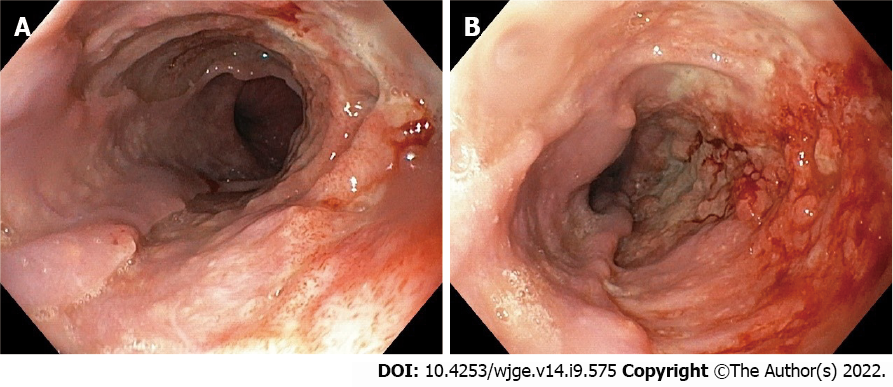
Figure 1 Upper gastrointestinal endoscopy.
A: Esophageal ulcer; B: Esophageal ulcer with nodules.
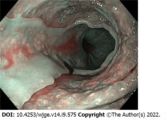
Figure 2 Esophageal biopsies.
Esophageal ulcer detected in narrow band imaging.
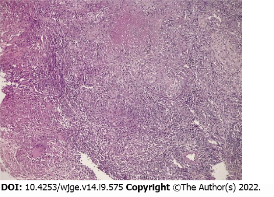
Figure 3 Granuloma with caseous necrosis (hematoxylin-eosin: 10 ×).
- Citation: Diallo I, Touré O, Sarr ES, Sow A, Ndiaye B, Diawara PS, Dial CM, Mbengue A, Fall F. Isolated esophageal tuberculosis: A case report. World J Gastrointest Endosc 2022; 14(9): 575-580
- URL: https://www.wjgnet.com/1948-5190/full/v14/i9/575.htm
- DOI: https://dx.doi.org/10.4253/wjge.v14.i9.575


