©The Author(s) 2022.
World J Gastrointest Endosc. Jun 16, 2022; 14(6): 402-415
Published online Jun 16, 2022. doi: 10.4253/wjge.v14.i6.402
Published online Jun 16, 2022. doi: 10.4253/wjge.v14.i6.402
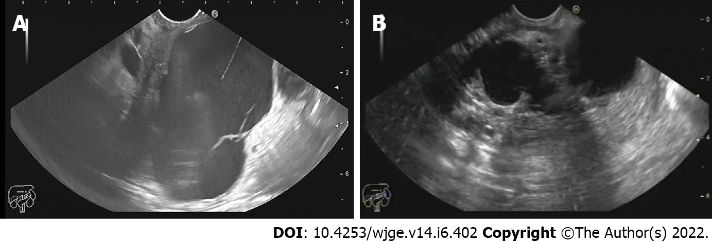
Figure 1 Pancreatic body mucinous cystadenoma.
A: Pancreatic body mucinous cystadenoma; B: Bilocular inflammatory pseudocyst in the gastric body.
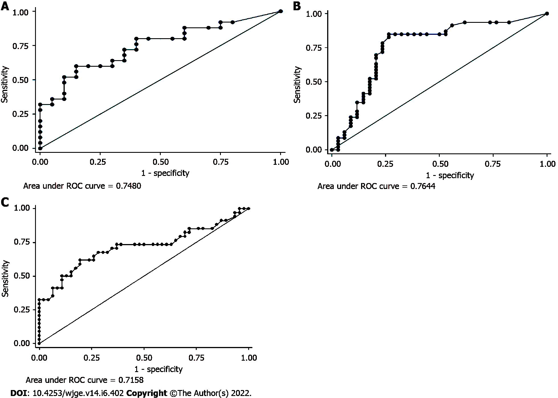
Figure 2 Receiver operating characteristic curve analysis.
A: Cyst fluid carcinoembryonic antigen level; B: Glucose level in cyst fluid; C: Cyst fluid serine protease inhibitor Kazal-type 1 level. ROC: Receiver operating characteristic.
- Citation: Okasha HH, Abdellatef A, Elkholy S, Mogawer MS, Yosry A, Elserafy M, Medhat E, Khalaf H, Fouad M, Elbaz T, Ramadan A, Behiry ME, Y William K, Habib G, Kaddah M, Abdel-Hamid H, Abou-Elmagd A, Galal A, Abbas WA, Altonbary AY, El-Ansary M, Abdou AE, Haggag H, Abdellah TA, Elfeki MA, Faheem HA, Khattab HM, El-Ansary M, Beshir S, El-Nady M. Role of endoscopic ultrasound and cyst fluid tumor markers in diagnosis of pancreatic cystic lesions. World J Gastrointest Endosc 2022; 14(6): 402-415
- URL: https://www.wjgnet.com/1948-5190/full/v14/i6/402.htm
- DOI: https://dx.doi.org/10.4253/wjge.v14.i6.402


