Copyright
©The Author(s) 2022.
World J Gastrointest Endosc. Jan 16, 2022; 14(1): 35-48
Published online Jan 16, 2022. doi: 10.4253/wjge.v14.i1.35
Published online Jan 16, 2022. doi: 10.4253/wjge.v14.i1.35
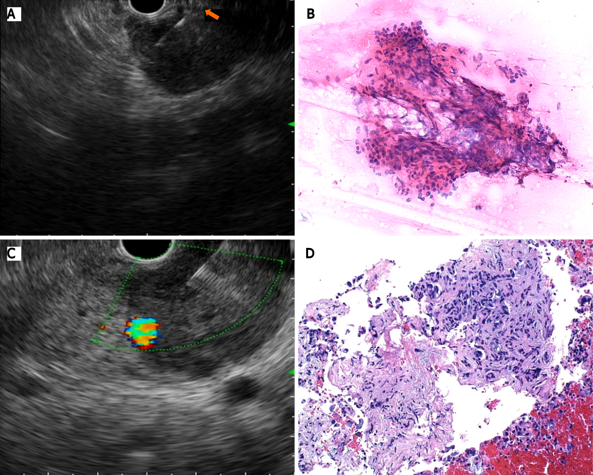
Figure 1 Endoscopic ultrasound-guided tisssue acquisition.
A: Puncture with a conventional fine needle aspiration needle; B: Pancreatic adenocarcinoma after cytologic evaluation; C: Tissue acquisition with a Franseen needle; D: Pancreatic tissue with preservation of cellular architecture.
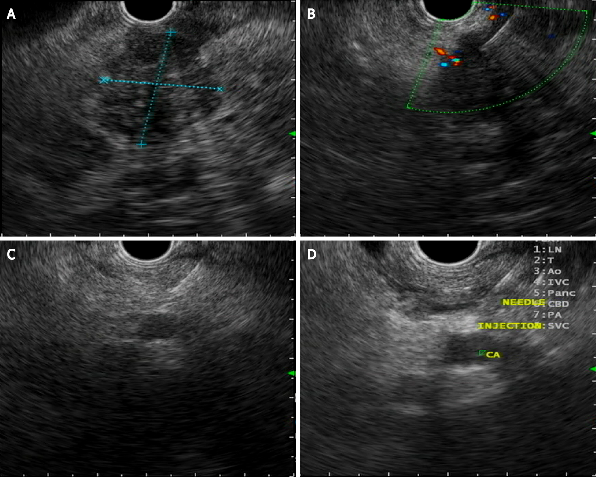
Figure 2 Celiac plexus neurolysis.
A: Pancreatic ductal adenocarcinoma located in the head of the pancreas; B: Endoscopic ultrasound (EUS)-guided tissue acquisition with a fine needle aspiration needle; C: EUS-guided puncture of the celiac plexus area; D: EUS-guided neurolysis with absolute alcohol injection.
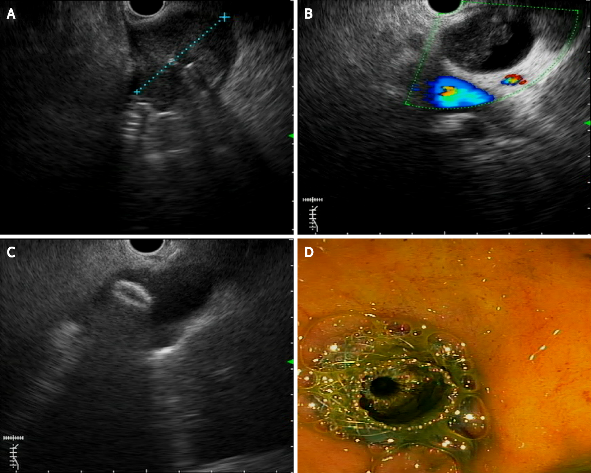
Figure 3 Endoscopic ultrasound-guided choledocoduodenostomy.
A: Pancreatic ductal adenocarcinoma (PDAC) located in the pancreatic head; B: Common bile duct dilation caused by PDAC; C: Lumen-appossable metallic stents (LAMS) distal flange opening inside the bile duct; D: Biliary drainage after LAMS placement.
- Citation: Salom F, Prat F. Current role of endoscopic ultrasound in the diagnosis and management of pancreatic cancer. World J Gastrointest Endosc 2022; 14(1): 35-48
- URL: https://www.wjgnet.com/1948-5190/full/v14/i1/35.htm
- DOI: https://dx.doi.org/10.4253/wjge.v14.i1.35


