©The Author(s) 2021.
World J Gastrointest Endosc. Sep 16, 2021; 13(9): 407-415
Published online Sep 16, 2021. doi: 10.4253/wjge.v13.i9.407
Published online Sep 16, 2021. doi: 10.4253/wjge.v13.i9.407
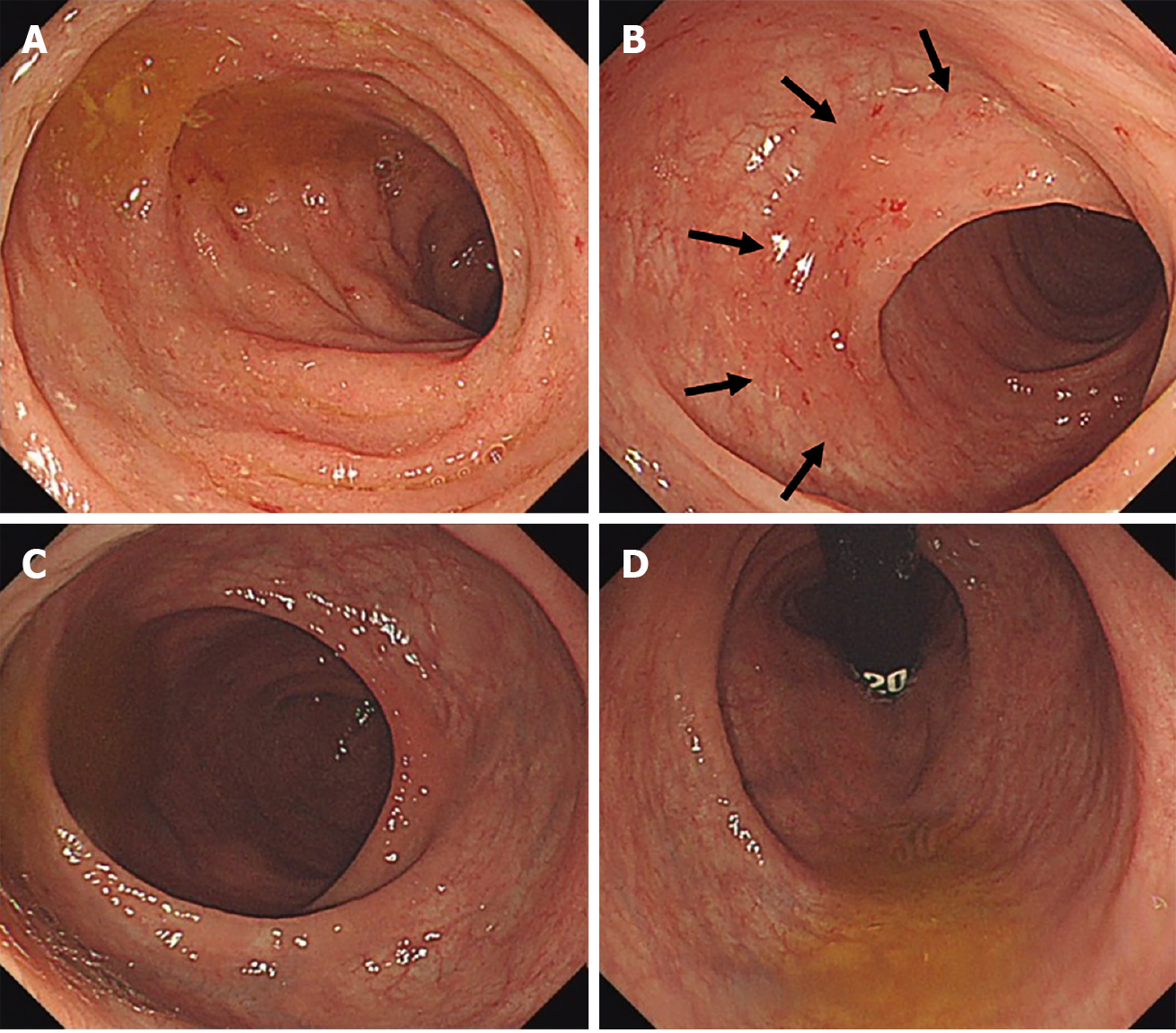
Figure 1 Colonoscopy at initial diagnosis.
A: On descending and sigmoid colon, continuous and symmetric micro-erosive inflammation with friability was noted; B: At distal sigmoid colon, transitional zone was noted (arrow); C: On the rectum, normal transparent mucosa with visible vascularity was noted; D: At retroflexion view, there was no evidence of mucosal inflammation.
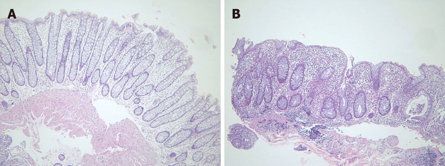
Figure 2 Hematoxylin and eosin stain.
A: Rectum: No architectural distortion or neutrophilic inflammation; B: Sigmoid colon: Crypt abscess, crypt distortion, and lymphoplasmacytic infiltration in lamina propria (hematoxylin and eosin stain × 100).
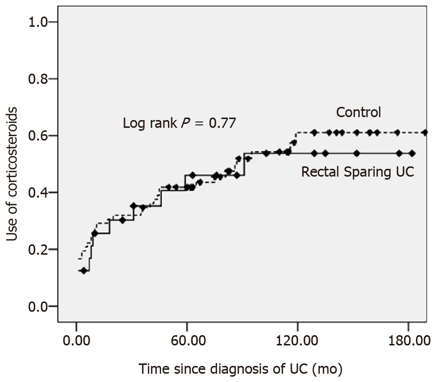
Figure 3 Cumulative rate of corticosteroids use in rectal sparing group (n = 24) vs control group (n = 72).
UC: Ulcerative colitis.
- Citation: Choi YS, Kim JK, Kim WJ. Clinical characteristics and prognosis of patients with ulcerative colitis that shows rectal sparing at initial diagnosis. World J Gastrointest Endosc 2021; 13(9): 407-415
- URL: https://www.wjgnet.com/1948-5190/full/v13/i9/407.htm
- DOI: https://dx.doi.org/10.4253/wjge.v13.i9.407


