©The Author(s) 2021.
World J Gastrointest Endosc. Aug 16, 2021; 13(8): 260-274
Published online Aug 16, 2021. doi: 10.4253/wjge.v13.i8.260
Published online Aug 16, 2021. doi: 10.4253/wjge.v13.i8.260
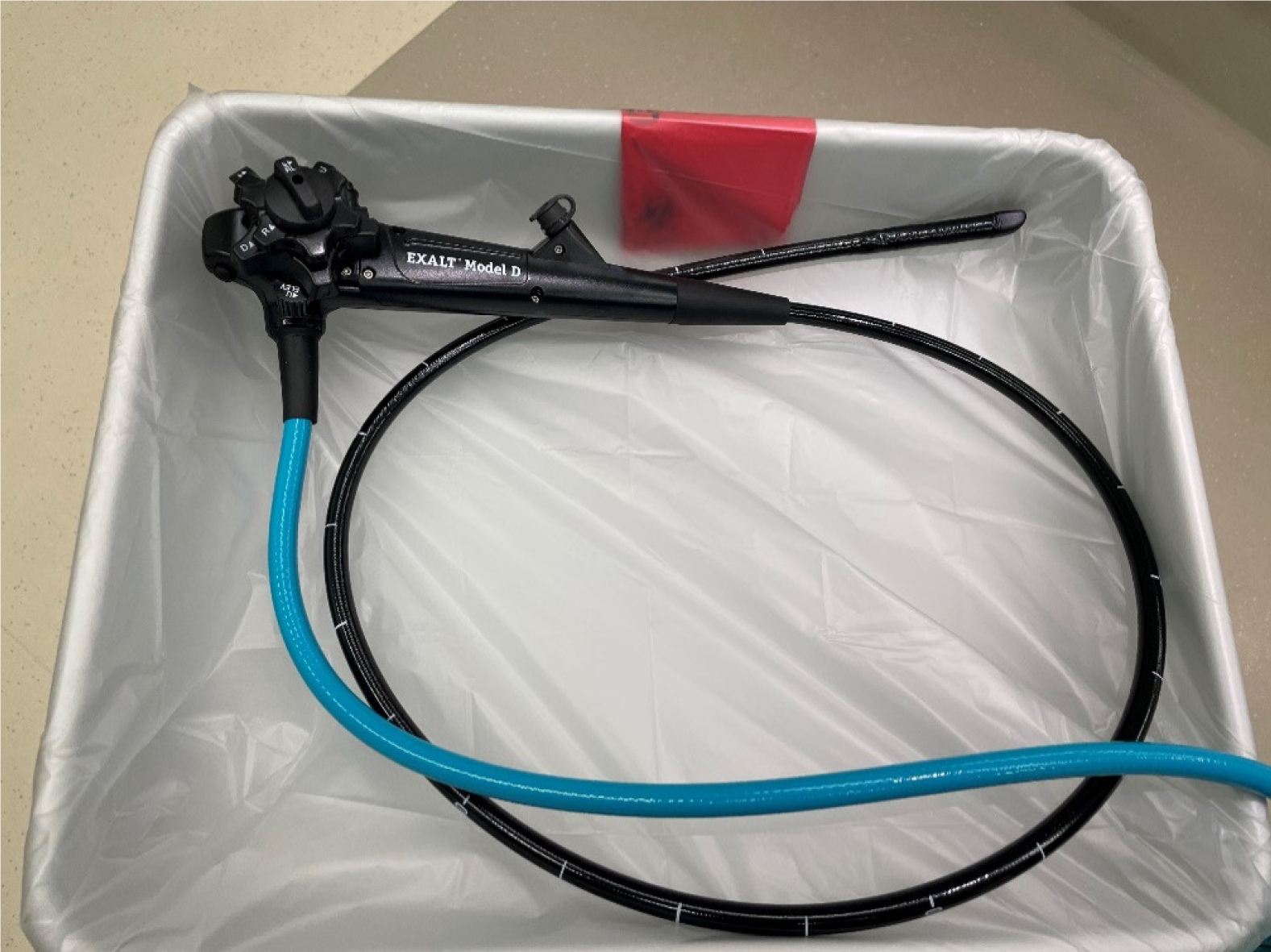
Figure 1 The EXALT duodenoscope in use at our center.
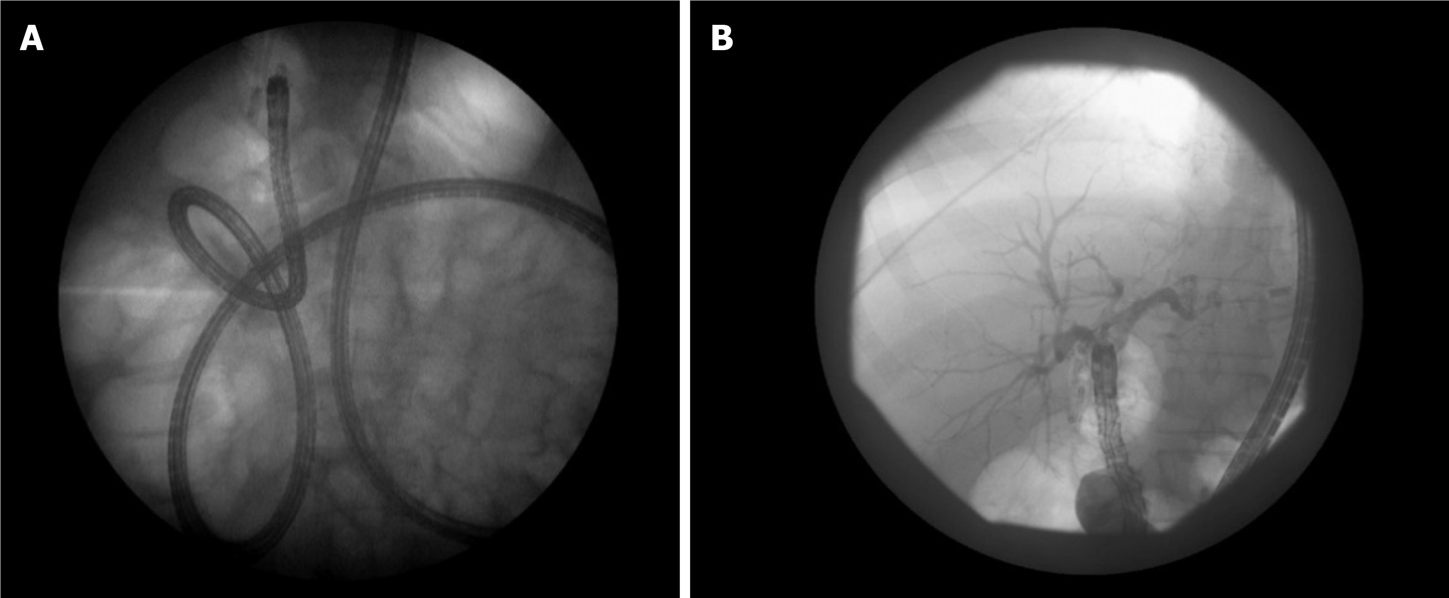
Figure 2 An overtube assisted enteroscopy and endoscopic retrograde cholangiopancreatography performed for a stent exchange and stone extraction.
The patient had a Roux-en-Y hepaticojejunostomy after a bile duct injury. A: Stent exchange; B: Stone extraction.
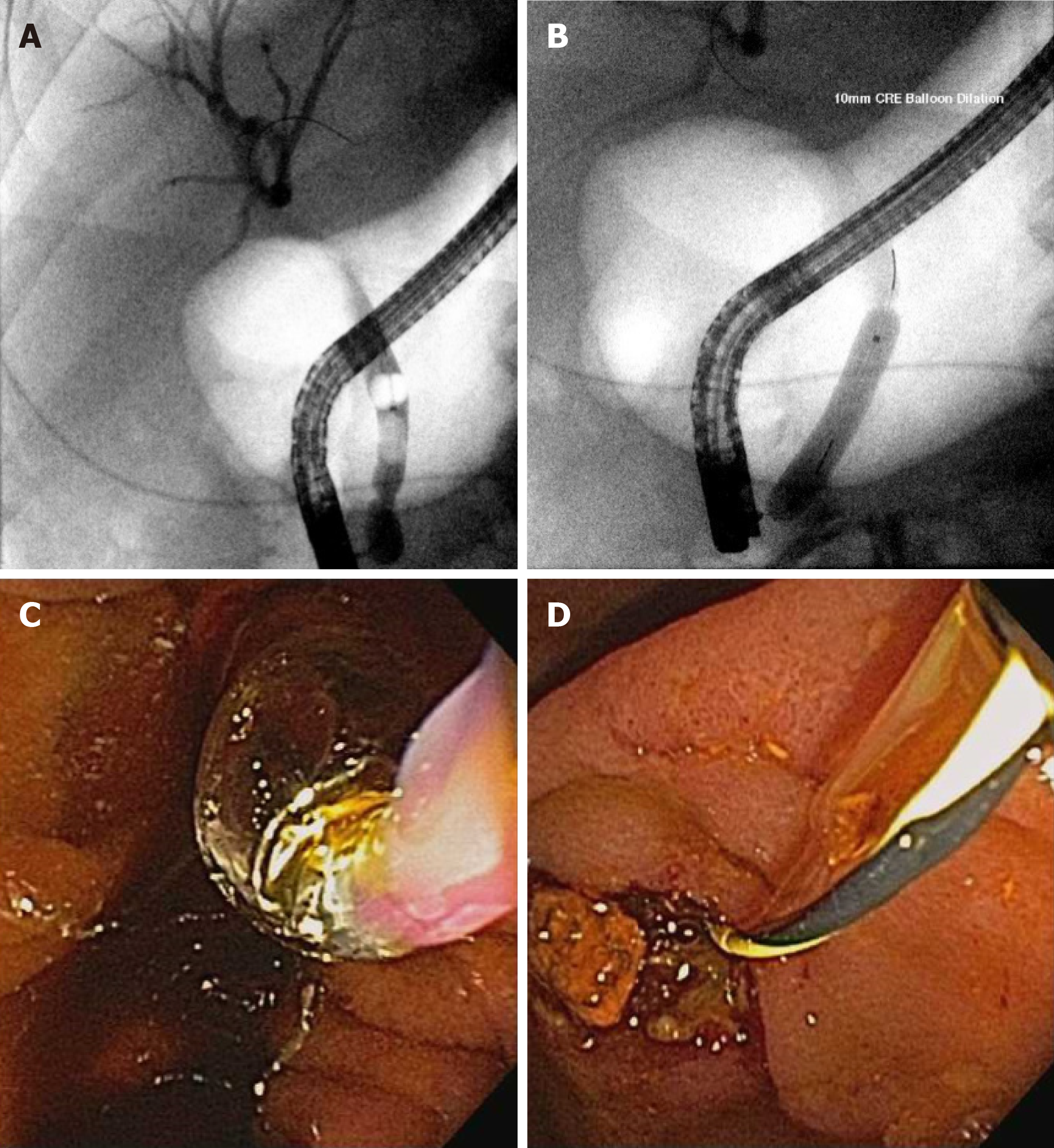
Figure 3 Large bile duct stone extraction.
A: Bile duct stone; B-D: Balloon sphincteroplasty performed (B and C) with extracted stone fragment (D).
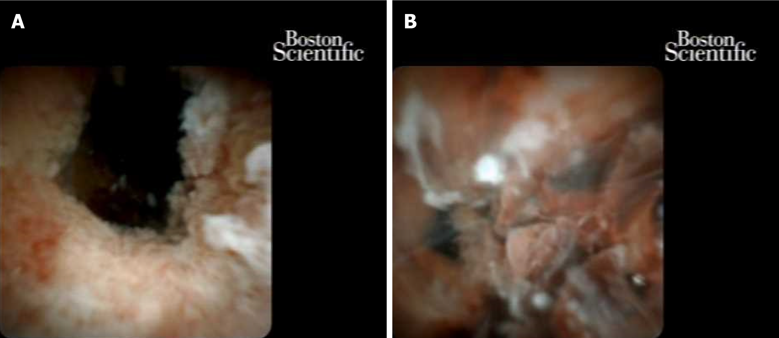
Figure 4 Cholangioscopy: Multifocal intraductal papillary neoplasm of bile ducts with high-grade dysplasia, that became cholangiocarcinoma.
A: High-grade dysplasia; B: Cholangiocarcinoma.
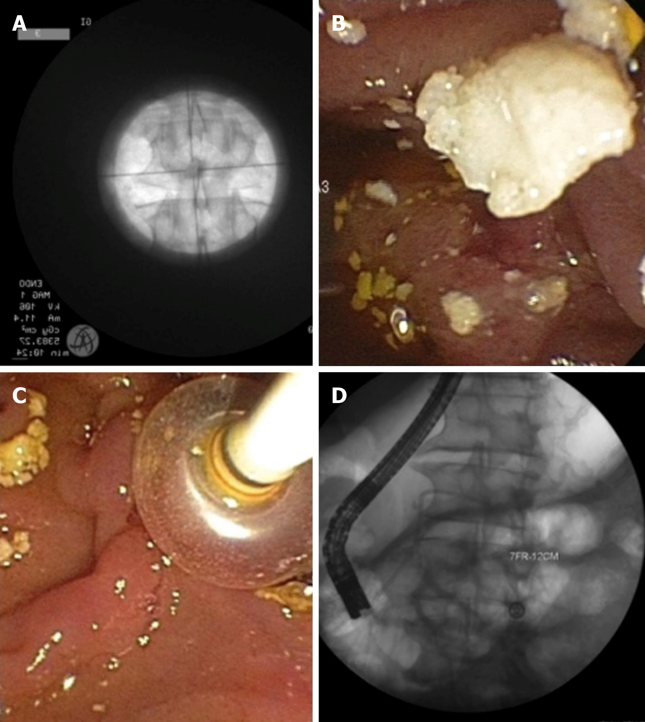
Figure 5 Chronic pancreatitis with a large pancreatic stone.
A: Extracorpeal shock wave lithotripsy with stone; B and C: Successful stone extraction; D: Placement of a plastic pancreatic stent.
- Citation: Sanders DJ, Bomman S, Krishnamoorthi R, Kozarek RA. Endoscopic retrograde cholangiopancreatography: Current practice and future research. World J Gastrointest Endosc 2021; 13(8): 260-274
- URL: https://www.wjgnet.com/1948-5190/full/v13/i8/260.htm
- DOI: https://dx.doi.org/10.4253/wjge.v13.i8.260


