©The Author(s) 2021.
World J Gastrointest Endosc. Nov 16, 2021; 13(11): 565-570
Published online Nov 16, 2021. doi: 10.4253/wjge.v13.i11.565
Published online Nov 16, 2021. doi: 10.4253/wjge.v13.i11.565
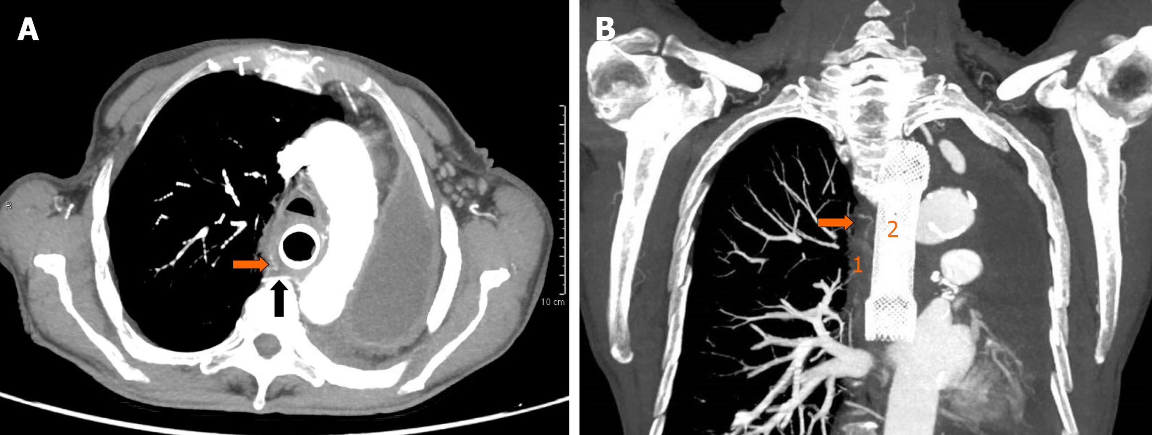
Figure 1 Arterial phase contrast-enhanced computed tomography.
A: Axial view showing the tortuous and dilated right bronchial artery (orange arrow) originating from the right third posterior intercostal artery (black arrow); B: Coronal view showing delation of the tissue planes between the right bronchial artery (orange arrow) and the thickened middle esophageal wall (1), with correct placement of the esophageal metal stent (2).
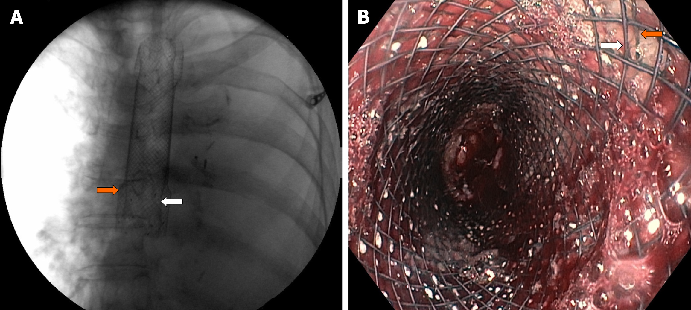
Figure 2 Placement of partially covered self-expandable metal stent (white arrow) through the previously inserted uncovered metal stent (orange arrow).
A: fluoroscopic view; B: Endoscopic view showing esophageal bleeding controlled by the partially covered metal stent.
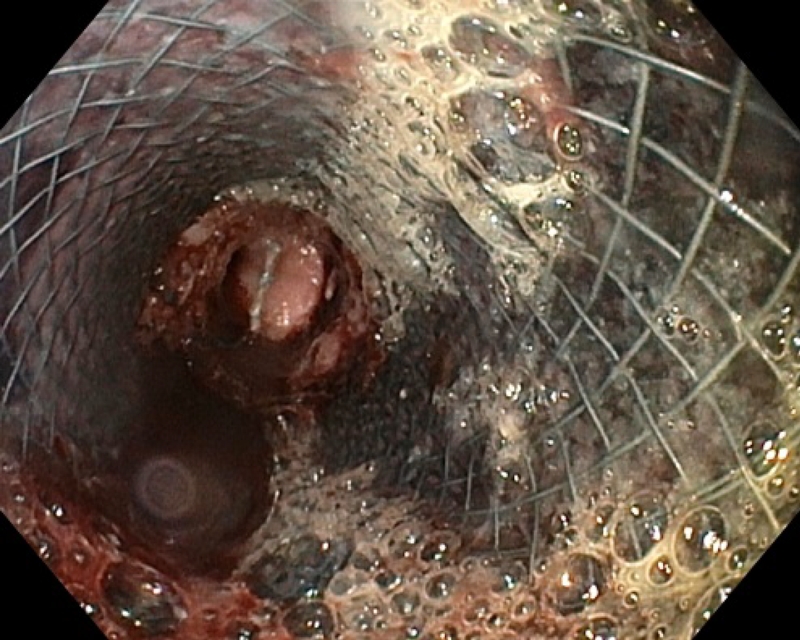
Figure 3
Second upper endoscopy showing fresh blood within the esophageal lumen and a diffuse amount of dark blood under the partially covered metal stent, in the absence of active bleeding sites.
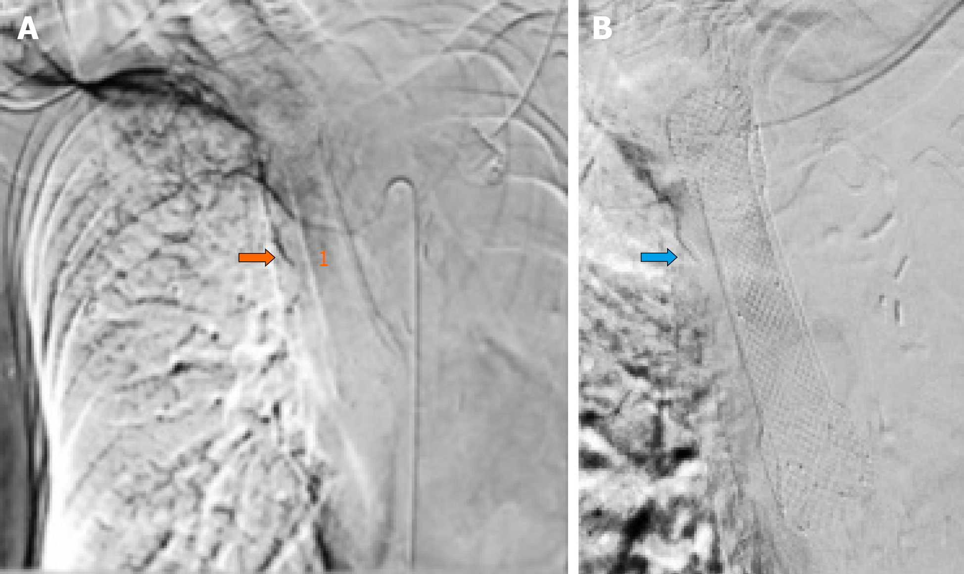
Figure 4 Operative angiography.
A: Selective arteriogram of the right bronchial artery (orange arrow) showing contrast extravasation within the esophageal lumen (1); B: Right bronchial artery coil embolization (blue arrow).
- Citation: Martino A, Oliva G, Zito FP, Silvestre M, Bennato R, Orsini L, Niola R, Romano L, Lombardi G. Acute upper gastrointestinal bleeding caused by esophageal right bronchial artery fistula: A case report. World J Gastrointest Endosc 2021; 13(11): 565-570
- URL: https://www.wjgnet.com/1948-5190/full/v13/i11/565.htm
- DOI: https://dx.doi.org/10.4253/wjge.v13.i11.565


