Copyright
©The Author(s) 2020.
World J Gastrointest Endosc. Oct 16, 2020; 12(10): 388-400
Published online Oct 16, 2020. doi: 10.4253/wjge.v12.i10.388
Published online Oct 16, 2020. doi: 10.4253/wjge.v12.i10.388
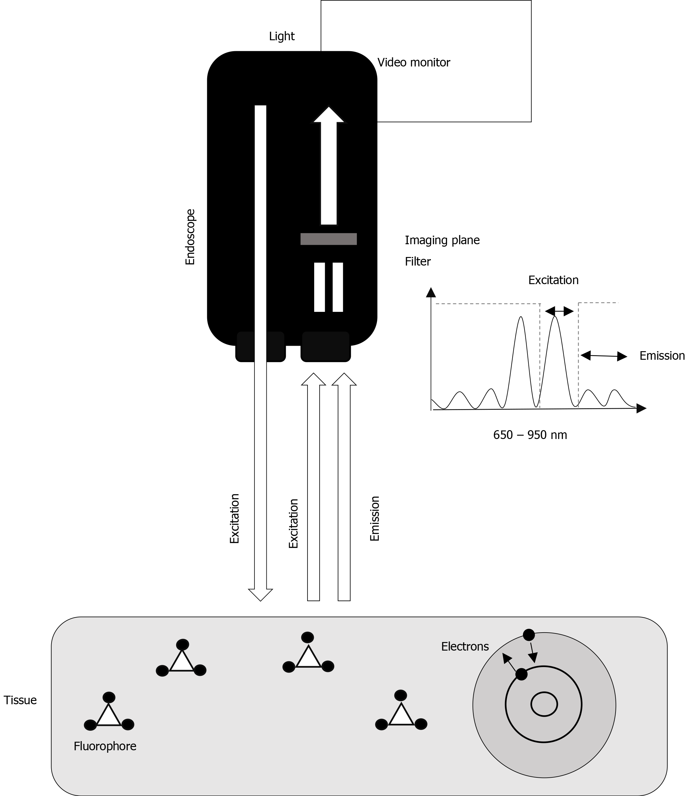
Figure 1 The endoscope emits light in the excitation spectrum of the fluorophore injected.
The electrons of the fluorophore will shift from one state of energy to another (excitation), and back, releasing energy as light (fluorescence) at another wavelength (emission). The imaging plane and the filter receive the signal and separate the signals of excitation and emission, only allowing the excitation light to pass.
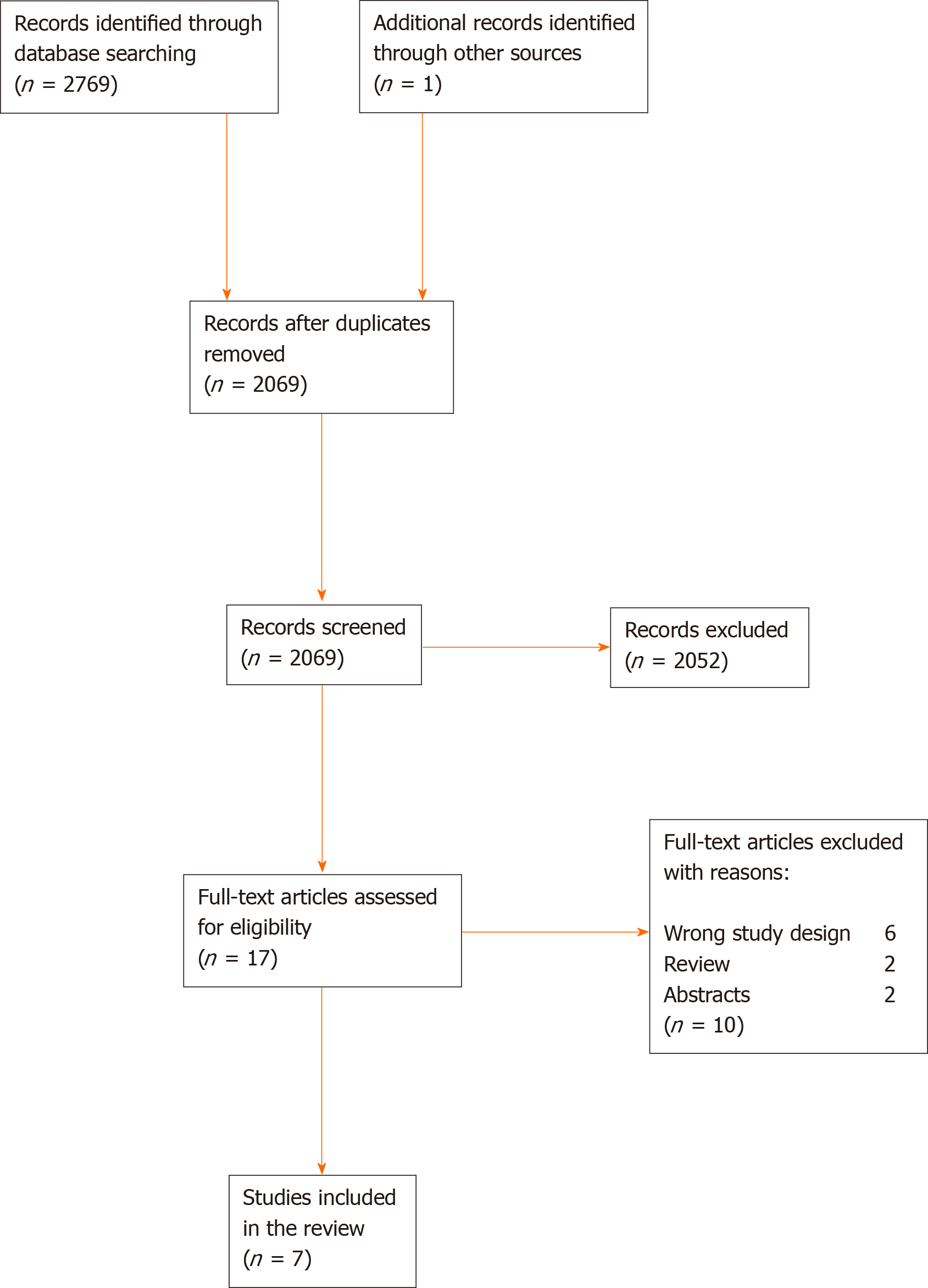
Figure 2 The screening process for the systematic review according to the PRISMA flow diagram.
- Citation: Mortensen OE, Nerup N, Thorsteinsson M, Svendsen MBS, Shiwaku H, Achiam MP. Fluorescence guided intraluminal endoscopy in the gastrointestinal tract: A systematic review. World J Gastrointest Endosc 2020; 12(10): 388-400
- URL: https://www.wjgnet.com/1948-5190/full/v12/i10/388.htm
- DOI: https://dx.doi.org/10.4253/wjge.v12.i10.388


