©The Author(s) 2020.
World J Gastrointest Endosc. Jan 16, 2020; 12(1): 23-32
Published online Jan 16, 2020. doi: 10.4253/wjge.v12.i1.23
Published online Jan 16, 2020. doi: 10.4253/wjge.v12.i1.23
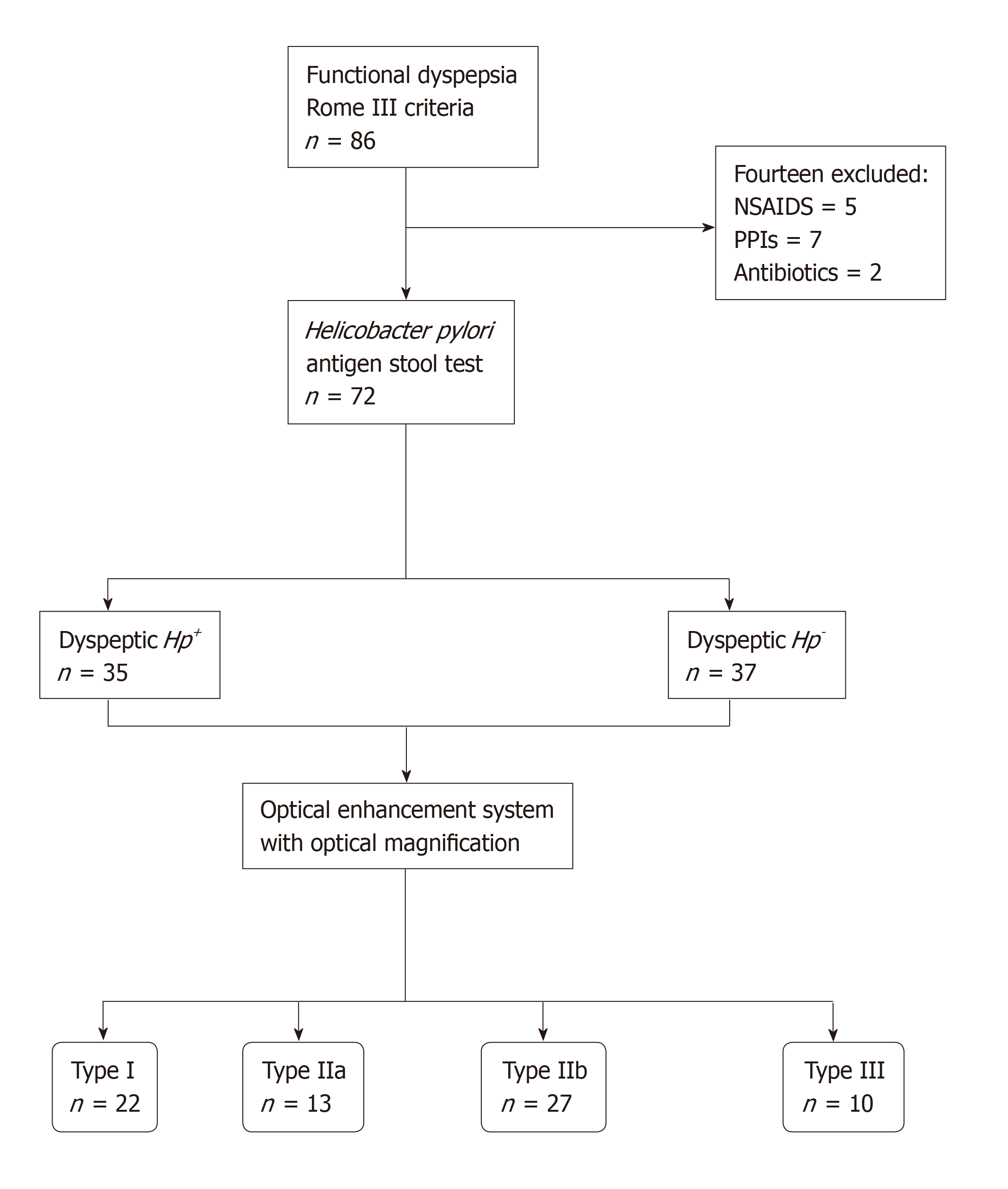
Figure 1 Flowchart showing the process of classification of the study participants.
NSAIDs: Nonsteroidal anti-inflammatory drugs; PPIs: Proton-pump inhibitors; Hp: Helicobacter pylori.
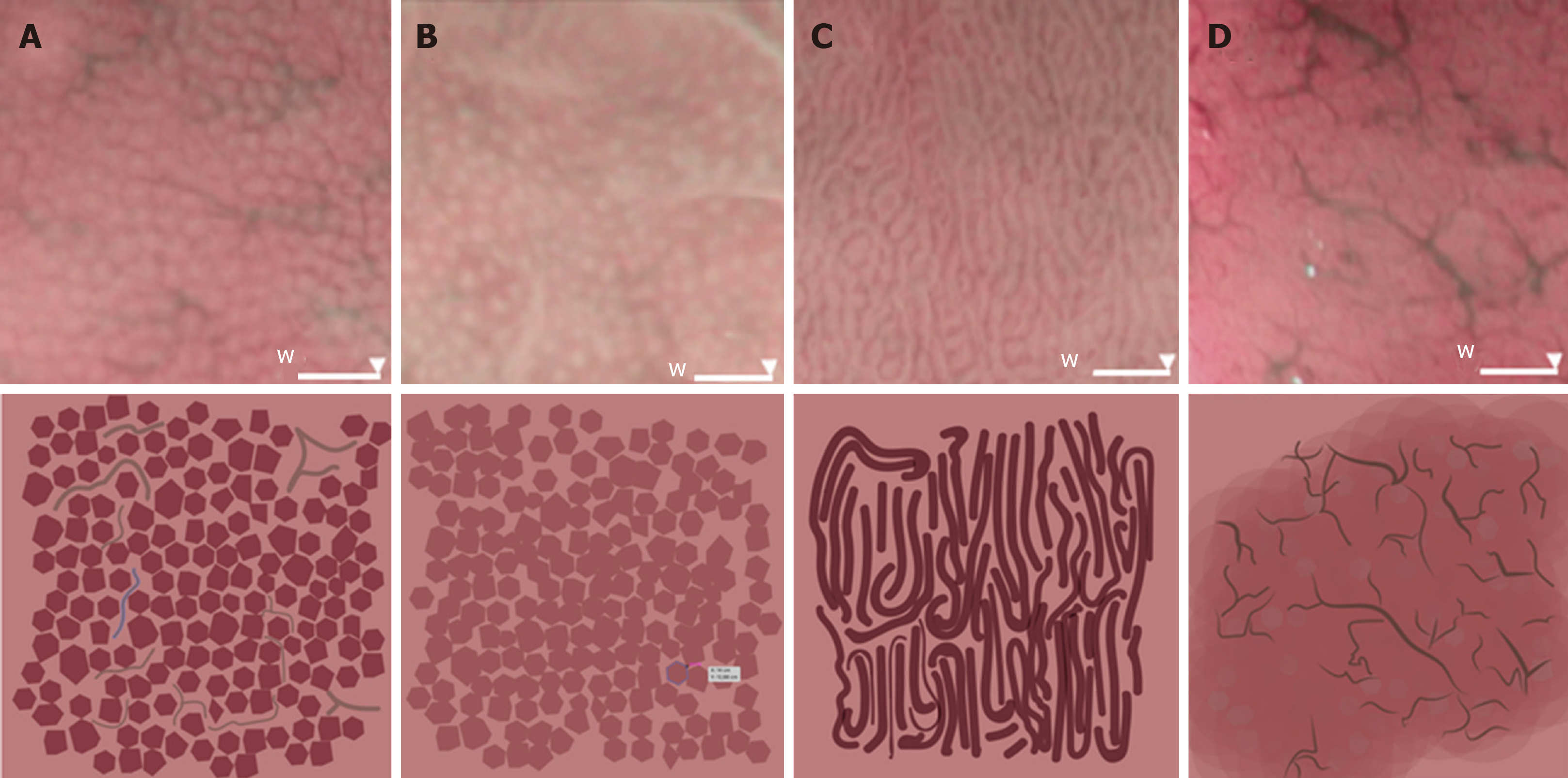
Figure 2 Four microvascular patterns identified by endoscopy of the gastric body mucosa.
A: The type I pattern comprises a honeycomb-type subepithelial capillary network (SECN), with a regular arrangement of collecting venules and regular, round pits; B: The type IIa pattern comprises a honeycomb-type SECN with regular, round pits, but with loss of collecting venules; C: In the type IIb pattern, there is loss of the normal SECN and collecting venules, and the presence instead of enlarged white pits surrounded by erythema; D: The type III pattern is characterized by loss of the normal SECN and round pits, with irregular arrangement of the collecting venules.
- Citation: Robles-Medranda C, Valero M, Puga-Tejada M, Oleas R, Baquerizo-Burgos J, Soria-Alcívar M, Alvarado-Escobar H, Pitanga-Lukashok H. High-definition optical magnification with digital chromoendoscopy detects gastric mucosal changes in dyspeptic-patients. World J Gastrointest Endosc 2020; 12(1): 23-32
- URL: https://www.wjgnet.com/1948-5190/full/v12/i1/23.htm
- DOI: https://dx.doi.org/10.4253/wjge.v12.i1.23


