©The Author(s) 2019.
World J Gastrointest Endosc. Feb 16, 2019; 11(2): 84-94
Published online Feb 16, 2019. doi: 10.4253/wjge.v11.i2.84
Published online Feb 16, 2019. doi: 10.4253/wjge.v11.i2.84
Figure 1 Overview of endoscopic surveillance of primary sclerosing cholangitis.
AASLD: American Association for the Study of Liver Diseases; CA 19-9: Carbohydrate antigen 19-9 serum tumor marker; EGD: Esophagogastroduodenoscopy; PSC: Primary sclerosing cholangitis; PSC-IBD: Inflammatory bowel disease co-existing with primary sclerosing cholangitis.
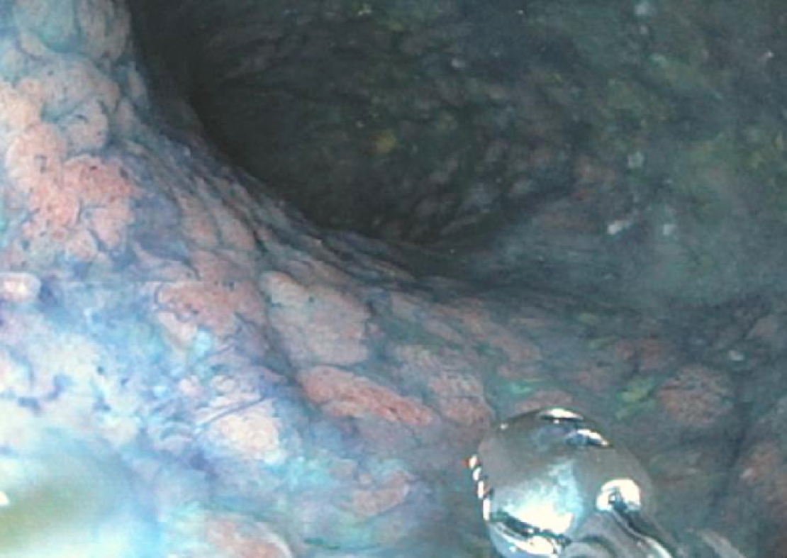
Figure 2 Example of the use of chromoendoscopy in a patient with primary sclerosing cholangitis.
In this example, the colon was irrigated using a methylene blue solution. Targeted biopsies were obtained in a region of the sigmoid colon where decreased uptake of methylene blue revealed a diffusely flat, nodular region. Pathology revealed multifocal low-grade dysplasia requiring total proctocolectomy.
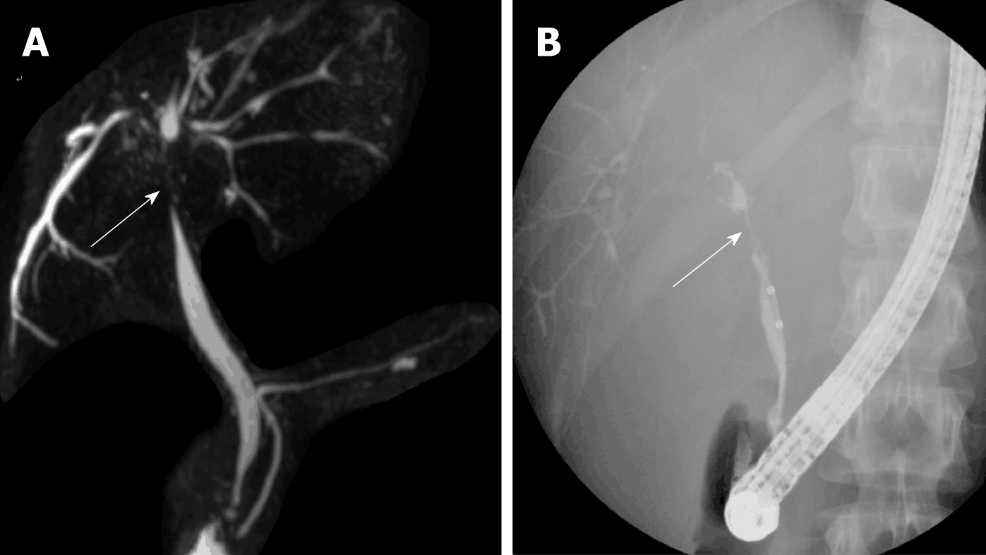
Figure 3 Examples of dominant strictures (arrow) in primary sclerosing cholangitis patients as seen on magnetic resonance cholangiopancreatography (A) and on endoscopic retrograde cholangiography (B).
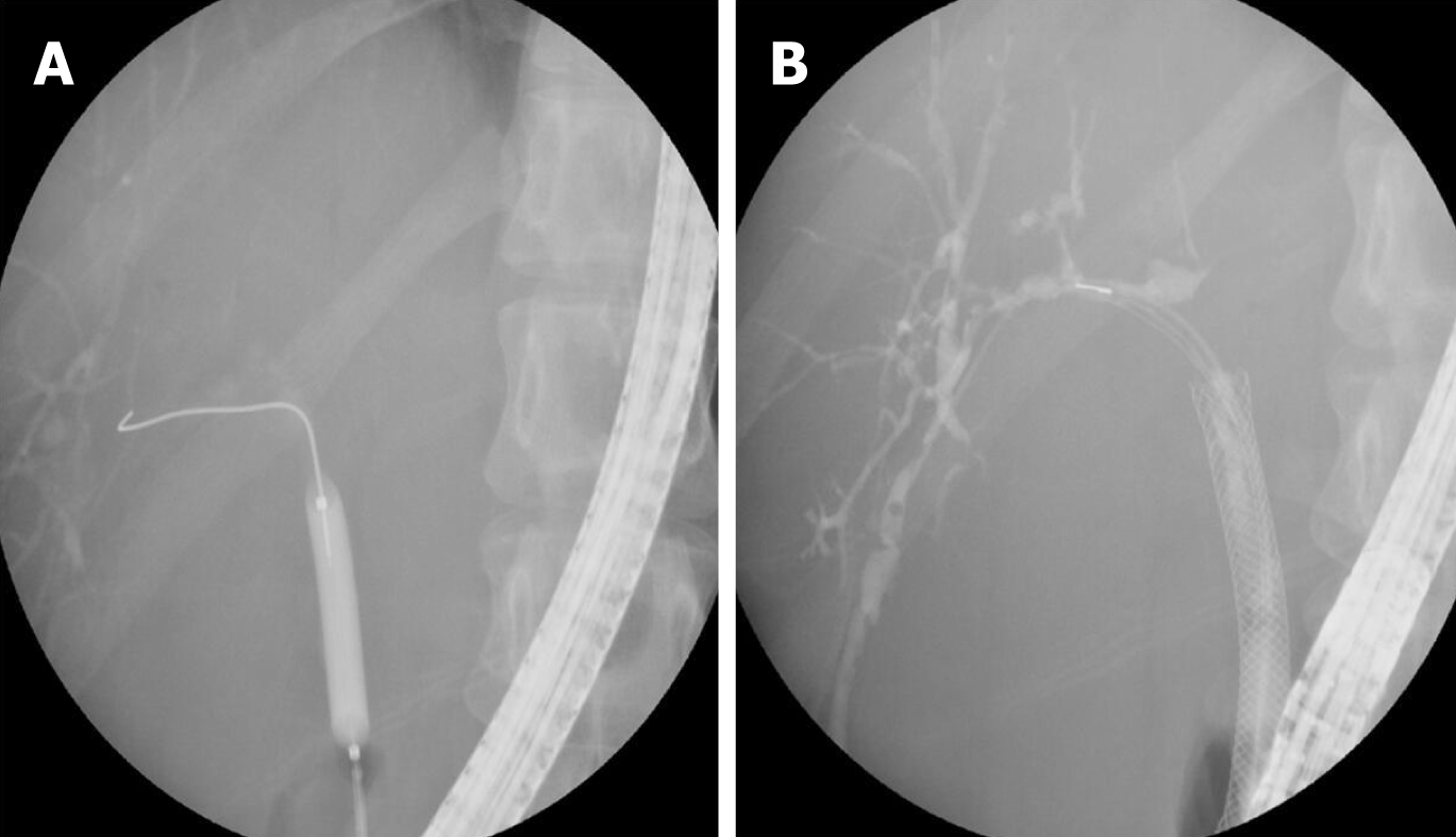
Figure 4 Examples of endoscopic management of a dominant stricture in a primary sclerosing cholangitis patient using balloon dilation (A) and short-term stenting (B).
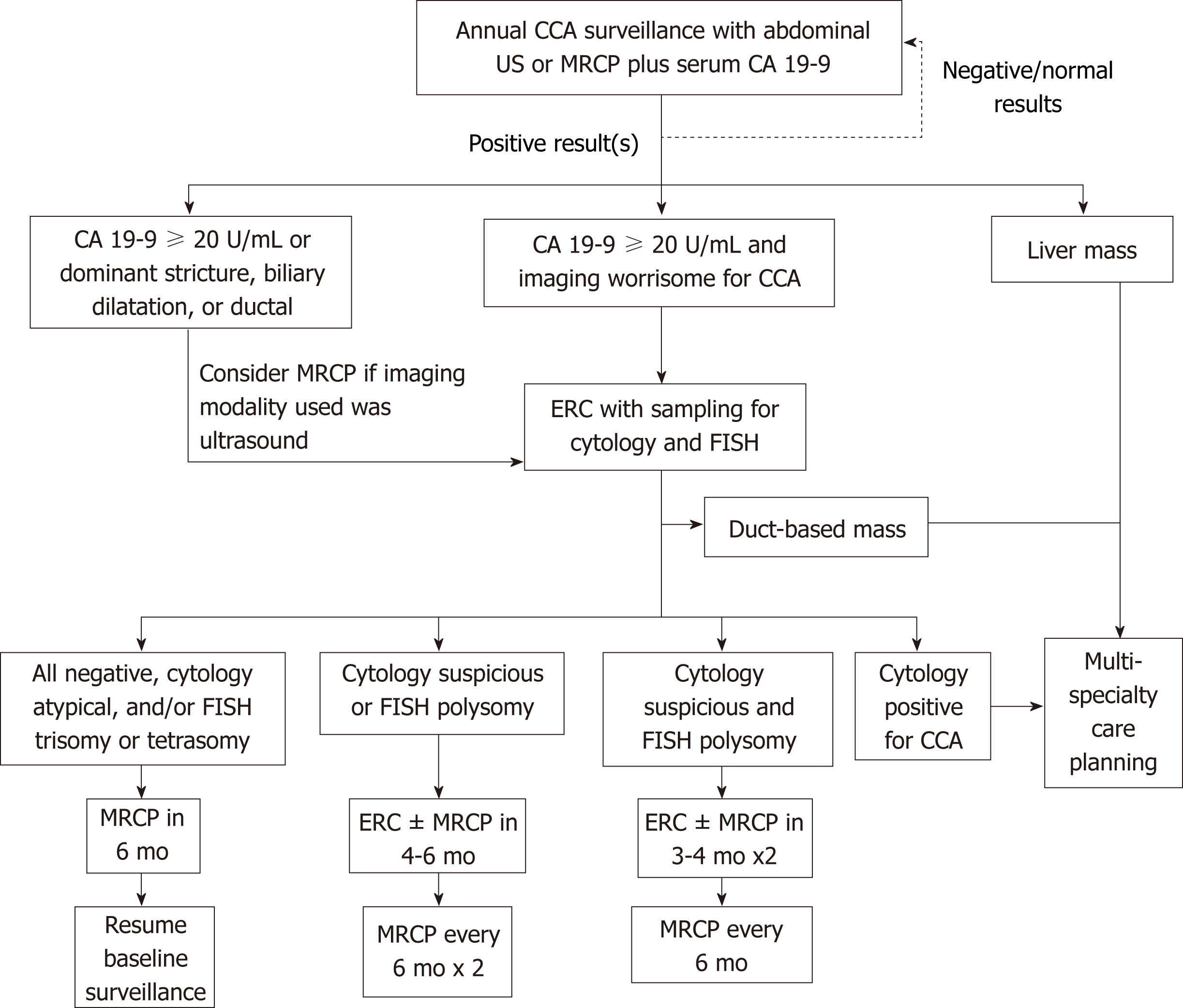
Figure 5 Proposed surveillance algorithm for cholangiocarcinoma in patients with primary sclerosing cholangitis.
CCA: Cholangiocarcinoma; US: Ultrasound; MRCP: Magnetic resonance cholangiopancreatography; CA 19-9: Carbohydrate antigen 19-9; ERC: Endoscopic retrograde cholaniography; FISH: Fluorescence in situ hybridization.
- Citation: Marya NB, Tabibian JH. Role of endoscopy in the management of primary sclerosing cholangitis. World J Gastrointest Endosc 2019; 11(2): 84-94
- URL: https://www.wjgnet.com/1948-5190/full/v11/i2/84.htm
- DOI: https://dx.doi.org/10.4253/wjge.v11.i2.84


