©The Author(s) 2018.
World J Gastrointest Endosc. Mar 16, 2018; 10(3): 69-73
Published online Mar 16, 2018. doi: 10.4253/wjge.v10.i3.69
Published online Mar 16, 2018. doi: 10.4253/wjge.v10.i3.69
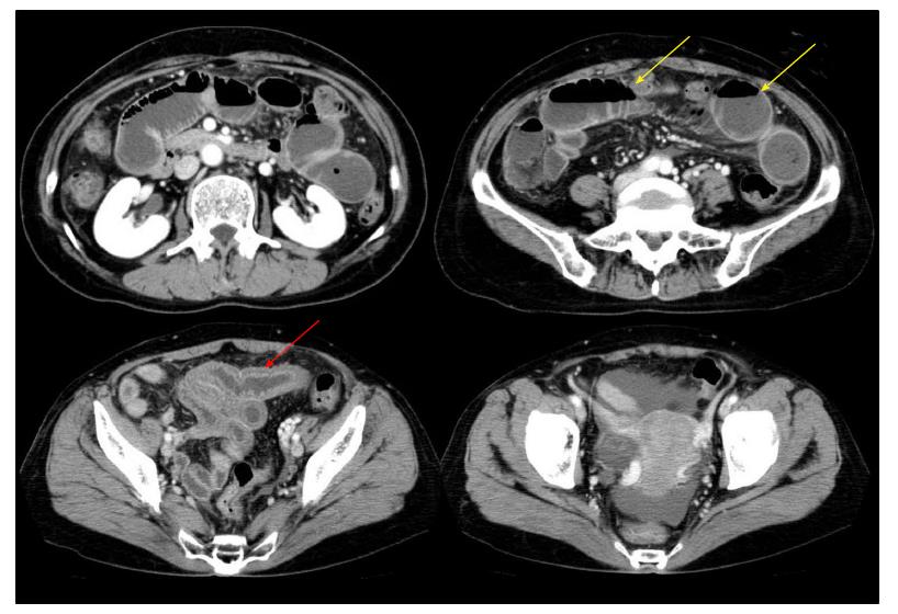
Figure 1 Abdominal computed tomography showing segmental edema of the intestinal wall (red arrow) with proximal dilatation (yellow arrow) and a small number of ascites.
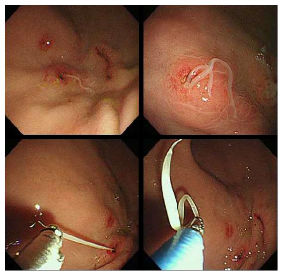
Figure 2 Anisakis larvae that are attached to the gastric mucosa can be removed using a biopsy forceps.
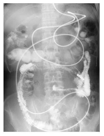
Figure 3 Ileography using gastrografin.
It did not reveal small intestine obstruction.
- Citation: Fujikawa H, Kuwai T, Yamaguchi T, Miura R, Sumida Y, Takasago T, Miyasako Y, Nishimura T, Iio S, Imagawa H, Yamaguchi A, Kouno H, Kohno H. Gastric and enteric anisakiasis successfully treated with Gastrografin therapy: A case report. World J Gastrointest Endosc 2018; 10(3): 69-73
- URL: https://www.wjgnet.com/1948-5190/full/v10/i3/69.htm
- DOI: https://dx.doi.org/10.4253/wjge.v10.i3.69


