©The Author(s) 2018.
World J Gastrointest Endosc. Dec 16, 2018; 10(12): 400-421
Published online Dec 16, 2018. doi: 10.4253/wjge.v10.i12.400
Published online Dec 16, 2018. doi: 10.4253/wjge.v10.i12.400
Figure 1 Flow diagrams - PRISMA[36].
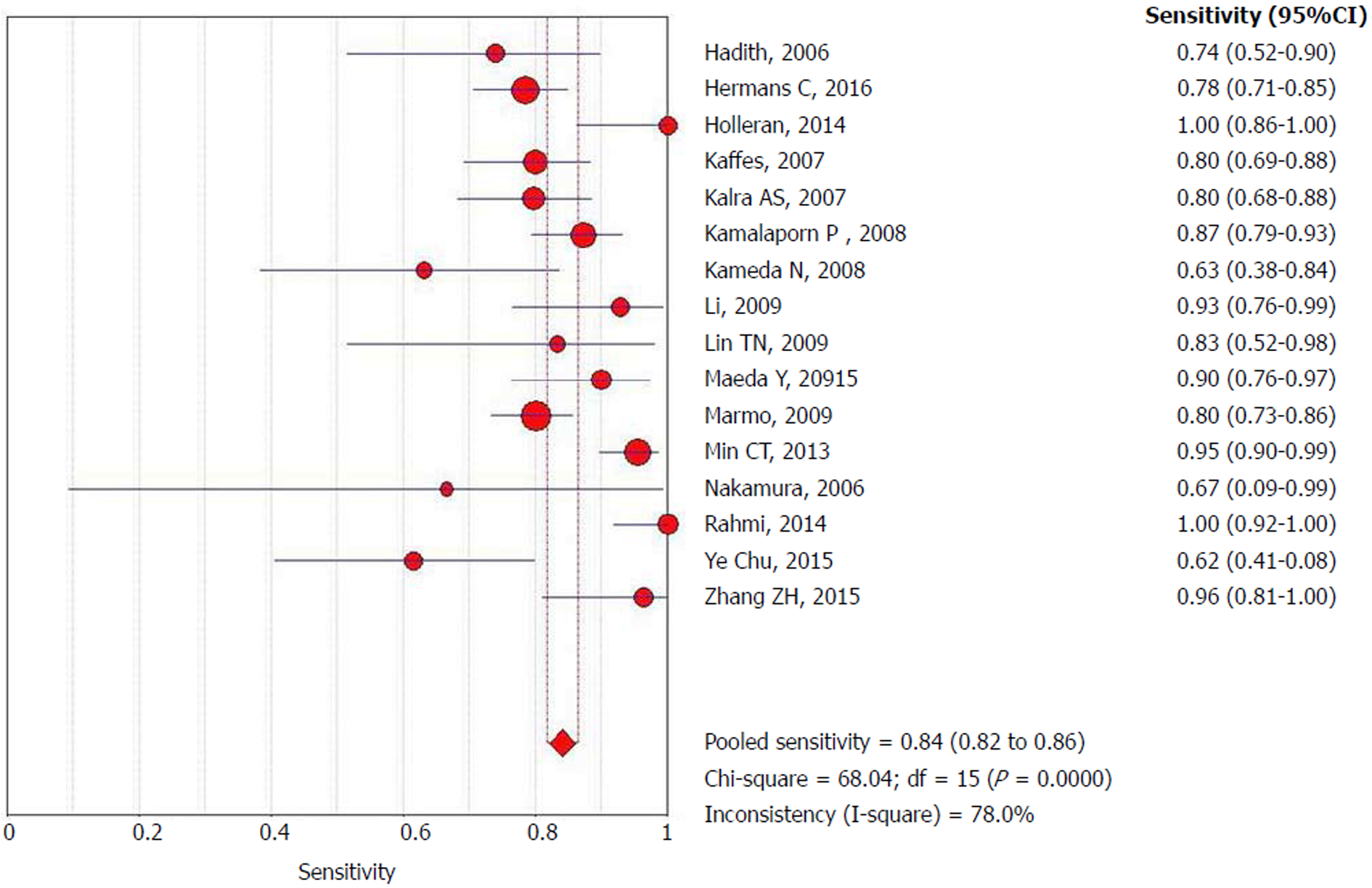
Figure 2 Forrest plot: Double-balloon enteroscopy sensitivity per-lesion analysis.
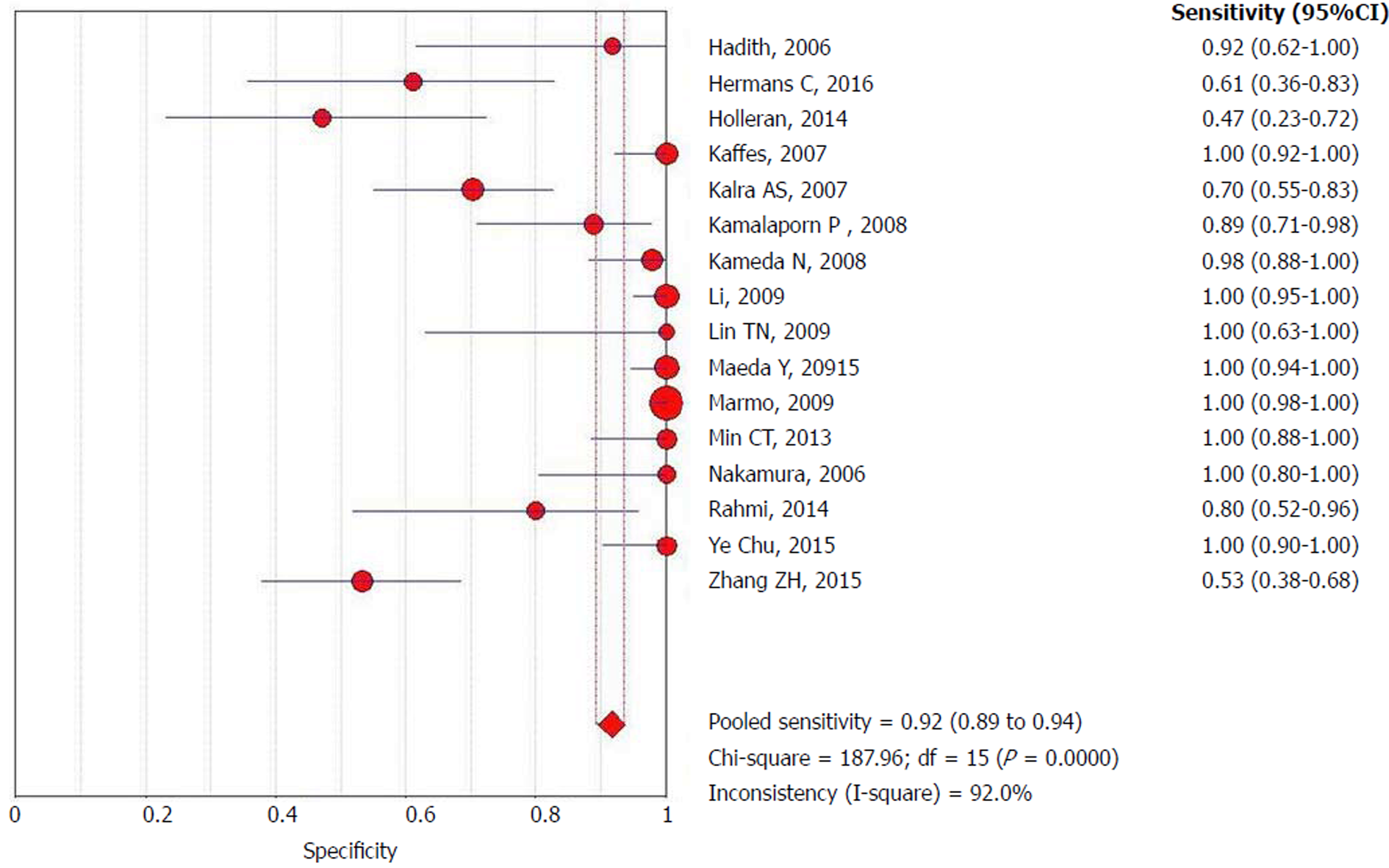
Figure 3 Forrest plot: Double-balloon enteroscopy specificity per-patient analysis.
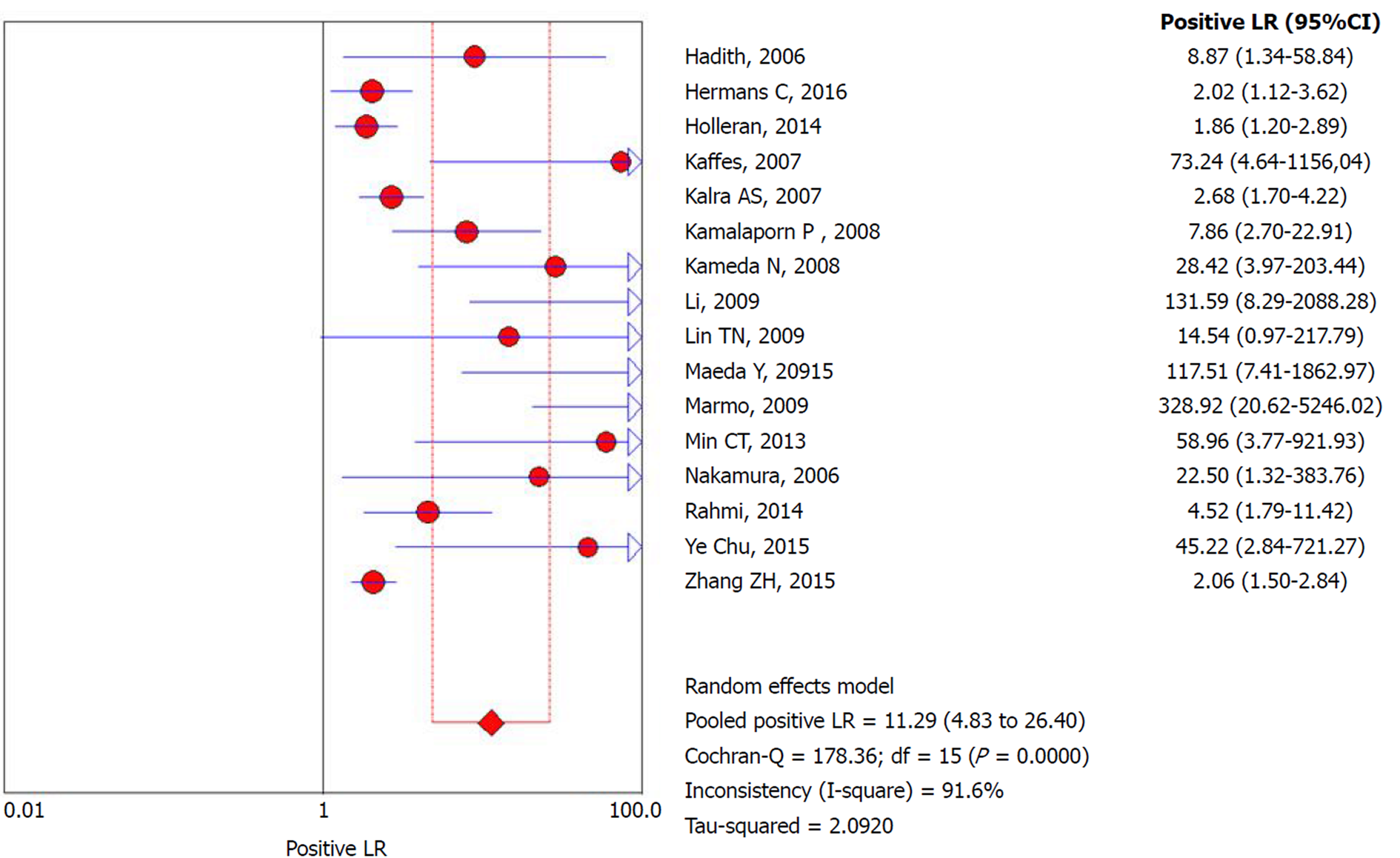
Figure 4 Forrest plot: Double-balloon enteroscopy positive likelihood ratio per-patient analysis.
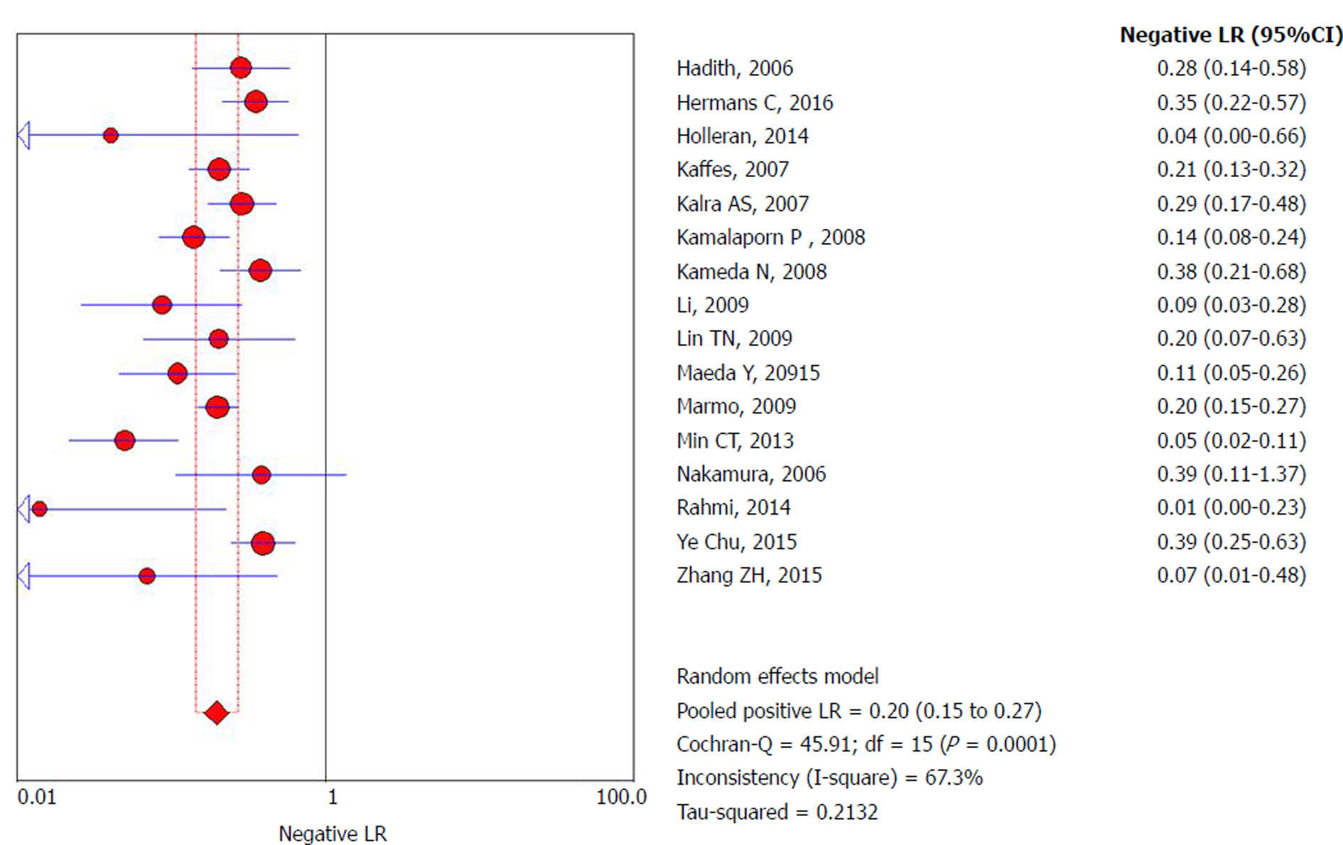
Figure 5 Forrest plot: Double-balloon enteroscopy negative likelihood ratio per-patient analysis.
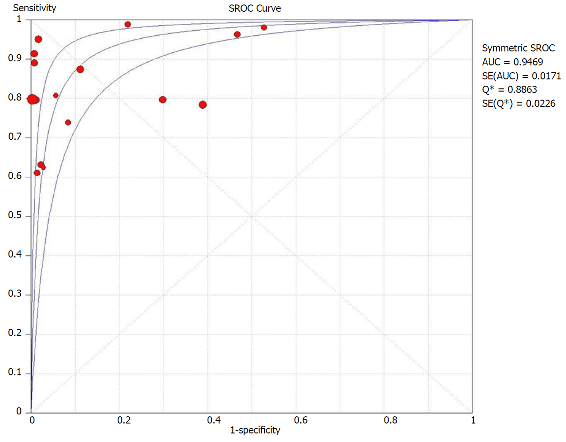
Figure 6 Summary receivers operating characteristic curve for double-balloon enteroscopy in per-patient analysis.
sROC: Summary receiver operating characteristic.
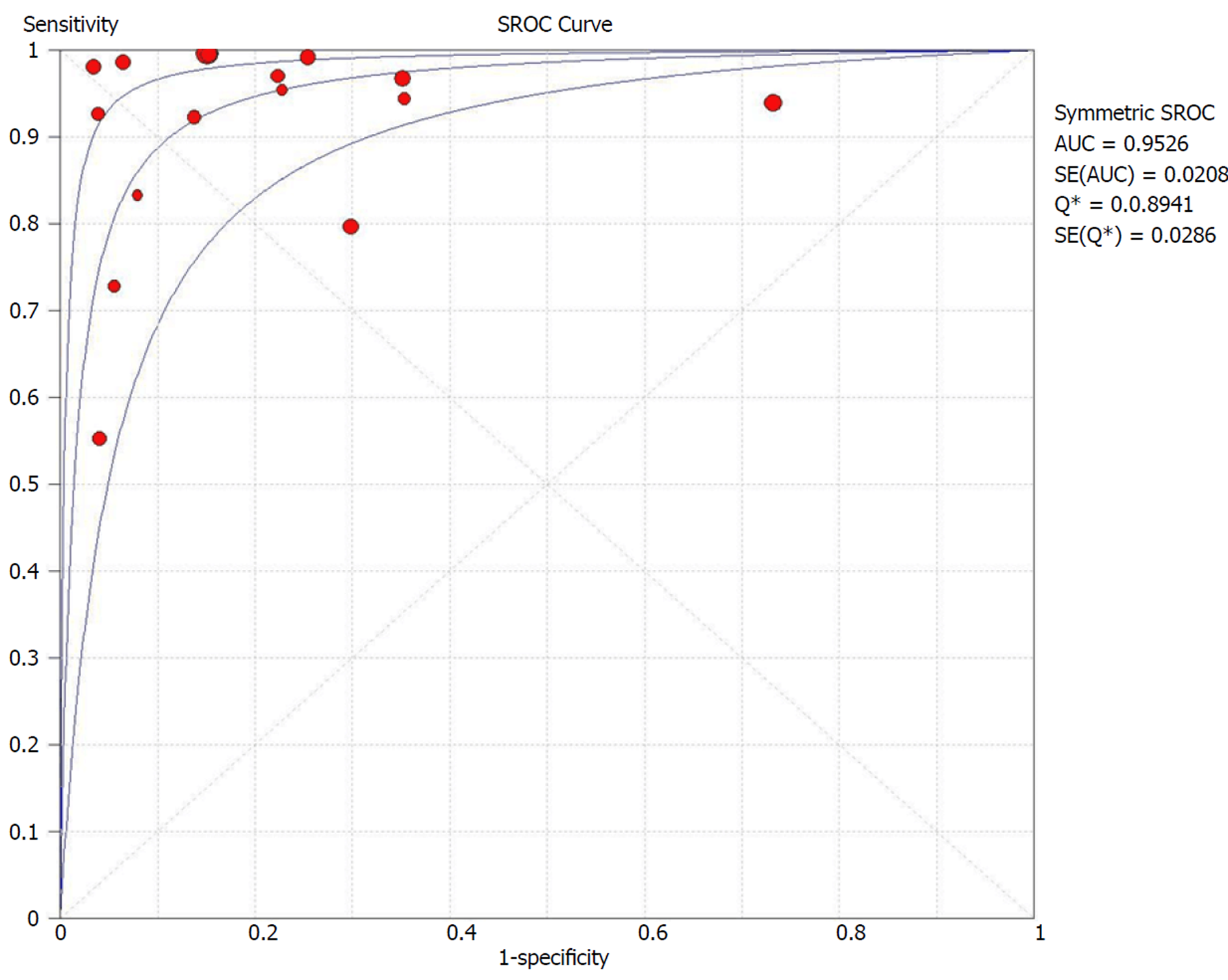
Figure 7 Summary receiver operating characteristic curve for video capsule endoscopy in per-patient analysis.
sROC: Summary receiver operating characteristic.
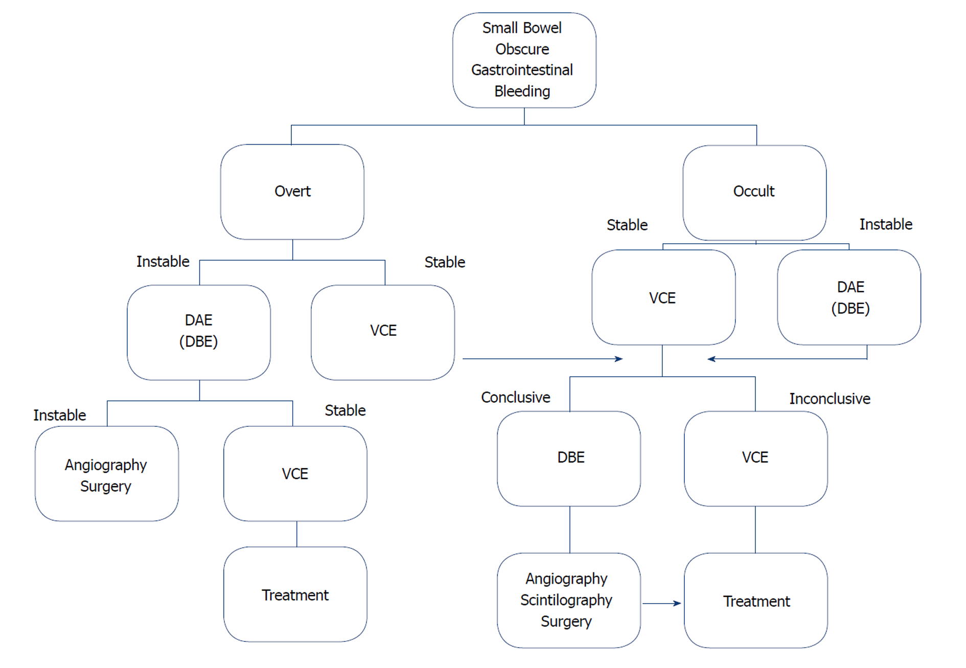
Figure 8 Suggested management approach to overt and occult small-bowel bleeding after upper endoscopy and colonoscopy did not identify vascular bleeding origin.
Positive test results should direct specific therapy. When video capsule endoscopy is contraindicated or unavailable, device-assisted endoscopy may serve as the initial test for small-bowel evaluation. VCE: Video capsule endoscopy; DAE: Device-assisted endoscopy; DBE: Double-balloon enteroscopy.
- Citation: Brito HP, Ribeiro IB, de Moura DTH, Bernardo WM, Chaves DM, Kuga R, Maahs ED, Ishida RK, de Moura ETH, de Moura EGH. Video capsule endoscopy vs double-balloon enteroscopy in the diagnosis of small bowel bleeding: A systematic review and meta-analysis. World J Gastrointest Endosc 2018; 10(12): 400-421
- URL: https://www.wjgnet.com/1948-5190/full/v10/i12/400.htm
- DOI: https://dx.doi.org/10.4253/wjge.v10.i12.400


