Copyright
©The Author(s) 2018.
World J Gastrointest Endosc. Oct 16, 2018; 10(10): 274-282
Published online Oct 16, 2018. doi: 10.4253/wjge.v10.i10.274
Published online Oct 16, 2018. doi: 10.4253/wjge.v10.i10.274
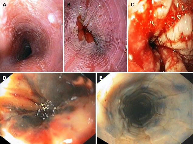
Figure 1 Modified Zargar's endoscopic classification of mucosal injury caused by ingestion of caustic substances.
A: Edema and erythema; B: Erosions and ulcers; C: Circumferential ulceration; D: Scattered areas of esophageal necrosis; E: Extensive esophageal necrosis.
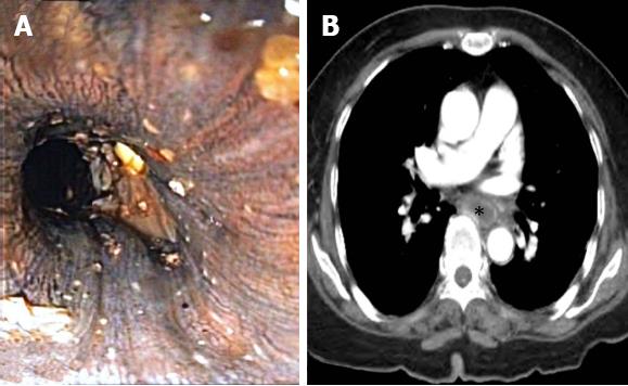
Figure 2 Endoscopic view suggested extensive mucosal necrosis of the esophagus -Grade IIIb modified Zargar's endoscopic classification, but CT scan revealed mucosal enhancement of the esophagus indicating tissue viability.
A: Endoscopic view; B: Computerized tomography scan. Notably, esophageal lumen is marked with asterisk.
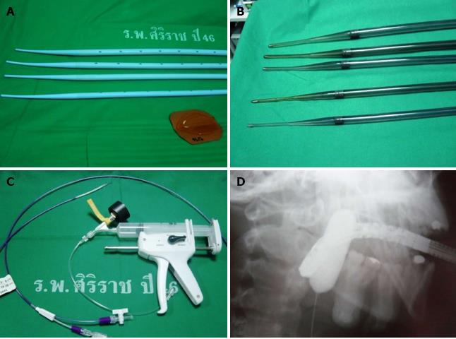
Figure 3 Various types of dilator.
A: Maloney-Hurst dilator; B: Savary-Gilliard dilator; C: Balloon dilator; D: Balloon dilator during dilatation seen with fluoroscopy.
- Citation: Methasate A, Lohsiriwat V. Role of endoscopy in caustic injury of the esophagus. World J Gastrointest Endosc 2018; 10(10): 274-282
- URL: https://www.wjgnet.com/1948-5190/full/v10/i10/274.htm
- DOI: https://dx.doi.org/10.4253/wjge.v10.i10.274


