©The Author(s) 2018.
World J Gastrointest Endosc. Jan 16, 2018; 10(1): 10-15
Published online Jan 16, 2018. doi: 10.4253/wjge.v10.i1.10
Published online Jan 16, 2018. doi: 10.4253/wjge.v10.i1.10
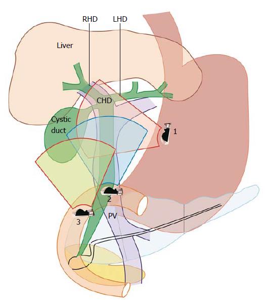
Figure 1 Station 1 shows the gall bladder at around 6 o’clock position; station 2 shows the gall bladder at around 3 o’clock position; and station 3 shows the gall bladder at around 9 o’clock position.
RHD: Right hepatic duct; LHD: Left hepatic duct; CHD: Common hepatic duct; PV: Portal vein.
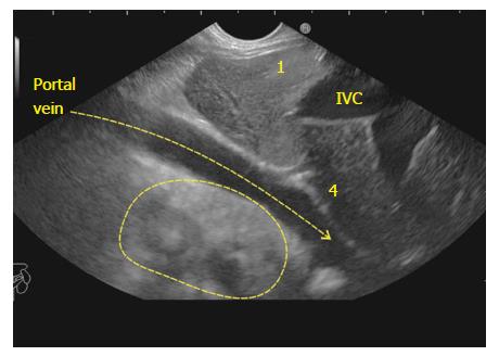
Figure 2 The supraduodenal part of portal vein is seen as a curving vessel going from 5/6 o’clock position to 9/10 o’clock position.
The yellow arrow points to the curving part of portal vein. The area marked with yellow outline shows the area in which the CBD and Gall Bladder can be seen. 1: Segment 1; 4: Segment 4; IVC: Inferior vena cava; CBD: Common bile duct.
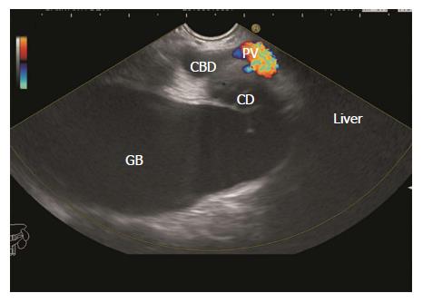
Figure 3 The upper part of common bile duct is first identified beyond the curving part of portal vein.
With slight rotation of the scope the cystic duct and gall bladder can be traced in the area beyond the portal vein between 5 o’clock position to 10 o’clock position. CBD: Common bile duct; GB: Gall bladder.
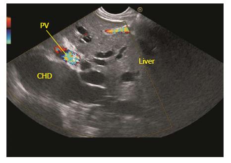
Figure 4 The dilated ducts of segment 2 and 3 can be followed to formation of left hepatic duct.
The left hepatic duct joins the right hepatic duct to form common hepatic duct. The common hepatic duct (CHD) lies beyond the supraduodenal part of portal vein. PV: Portal vein.
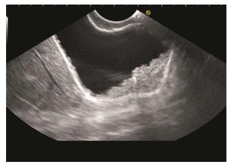
Figure 5 The gall bladder imaging is done from duodenal bulb.
The layers of GB can be seen. The irregular polypoidal mass occupying the lumen is due to adenomyomatosis of GB. GB: Gall bladder.
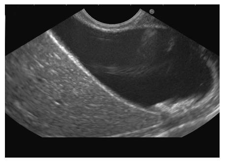
Figure 6 Gall bladder imaging from the duodenal bulb.
The stones are present in the lumen of GB. The neck of the Gall Bladder is present at 11 o’clock position and the fundus is present at 3 o’clock position. GB: Gall bladder.
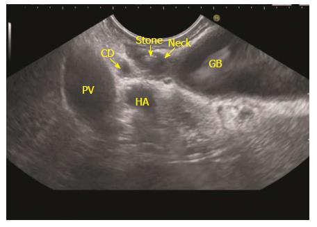
Figure 7 A stone is seen in the neck of gall bladder.
These stones can be missed by routine abdominal ultrasound. The neck of the gall bladder is present just below the probe and the fundus is present at 3 o’clock position. PV: Portal vein; GB: Gall bladder.
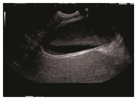
Figure 8 The segment 5 of liver is seen beyond the gall bladder.
A layer of gall bladder (GB) sludge is seen in the lumen of GB.
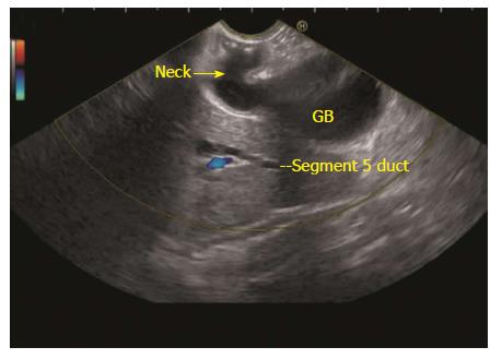
Figure 9 The neck of the gall bladder is present just below the probe and the fundus is present at 3 o’clock position.
The segment 5 duct is seen beyond the GB. GB: Gall bladder.
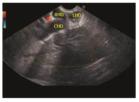
Figure 10 Once the gall bladder imaging is done from duodenal bulb an anticlockwise rotation can trace the common bile duct towards the hilum of liver.
The CHD is seen to be dividing into right and left hepatic duct. RHD: Right hepatic duct; LHD: Left hepatic duct; CHD: Common hepatic duct.
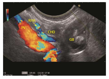
Figure 11 The imaging is done from duodenal bulb and the portal vein is identified going from 5 o’clock position to 10 o’clock position in a long axis.
The CHD is identified between the probe and portal vein. The CHD is followed up by anticlockwise rotation and the remnant of gall bladder is seen in continuity with CHD. CHD: Common hepatic duct; GB: Gall bladder; PV: Portal vein.
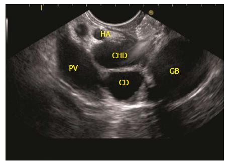
Figure 12 The imaging is done from duodenal bulb and the portal vein is identified going from 5 o’clock position to 10 o’clock position in a long axis.
The CHD is identified between the probe and portal vein. The CHD is followed up by anticlockwise rotation and the continuity into cystic duct and gall bladder is seen. CHD: Common hepatic duct; PV: Portal vein; GB: Gall bladder.
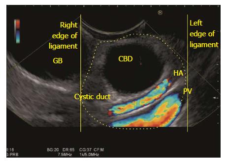
Figure 13 The gall bladder imaging is done from descending duodenum with up deflection and anti-clockwise rotation.
The hepatoduodenal ligament is identified as a bean shaped structure between the probe and liver (shown in dotted yellow area). The CBD can be traced along the cystic duct and the gall bladder which lies outside the right edge of hepatoduodenal ligament. CBD: Common bile duct; GB: Gall bladder.
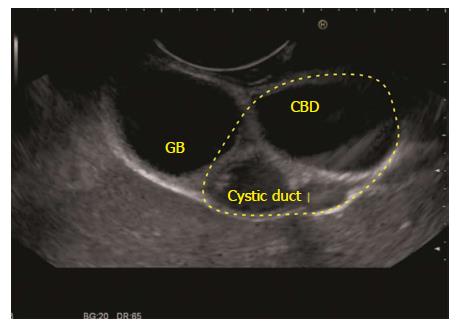
Figure 14 The gall bladder imaging is done from descending duodenum.
The hepatoduodenal ligament is identified between the probe and liver (shown in dotted yellow area). The CBD, the cystic duct and the gall bladder are visualized on the under surface of liver. CBD: Common bile duct; GB: Gall bladder.
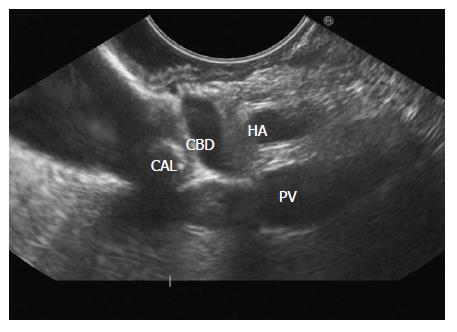
Figure 15 The gall bladder imaging is done from descending duodenum with up deflection and anti-clockwise rotation.
The CBD can be traced and a stone is seen in the Cystic duct. The distended gall bladder is also visualized. PV: Portal vein; CBD: Common bile duct.
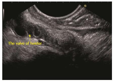
Figure 16 The gall bladder imaging is done from descending duodenum with up deflection and anti-clockwise rotation.
The tortuous cystic duct with a spiral valve of Heister is seen.
- Citation: Sharma M, Somani P, Sunkara T. Imaging of gall bladder by endoscopic ultrasound. World J Gastrointest Endosc 2018; 10(1): 10-15
- URL: https://www.wjgnet.com/1948-5190/full/v10/i1/10.htm
- DOI: https://dx.doi.org/10.4253/wjge.v10.i1.10


