©2014 Baishideng Publishing Group Inc.
World J Stem Cells. Sep 26, 2014; 6(4): 432-440
Published online Sep 26, 2014. doi: 10.4252/wjsc.v6.i4.432
Published online Sep 26, 2014. doi: 10.4252/wjsc.v6.i4.432
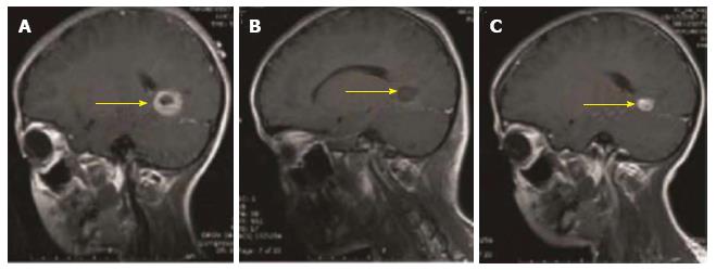
Figure 1 Magnetic resonance imaging graphs illustrate the presence, removal, and reappearance of a glioblastoma patient (yellow arrow: tumor mass).
A: Pre-operation, visualizing the presence of the tumor; B: Post-surgery, visualizing disappearance of the tumor; C: 3-mo post-surgery, visualizing the reappearance of the tumor.
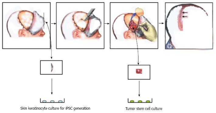
Figure 2 Personalized treatment of brain tumors by using autologous stem cells (induced pluripotent stem cells) through the induced pluripotent stem cells strategy for treating brain tumors.
During surgery, a piece of skin is obtained to generate induced pluripotent stem cells (iPSCs) while tumor cells are processed to obtain tumor stem cells (TSCs). The iPSCs are used to take therapy specific to autologous TSCs.
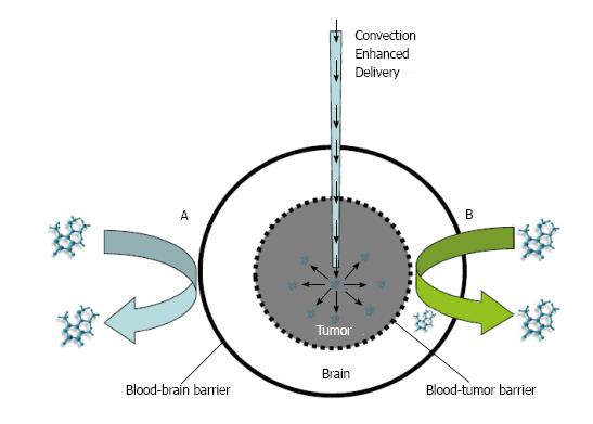
Figure 3 Convection enhanced delivery of therapy to overcome two barriers of brain tumors.
A: Systemic delivery of drugs blocked from entry into the brain by the blood brain barrier; B: Drug delivery inhibited by the brain-tumor barrier. This convection enhanced delivery can be used to deliver neural stem cells locally onto a tumor.
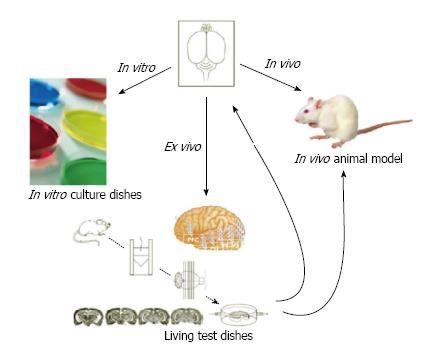
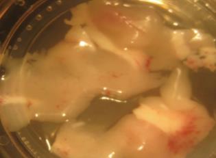
Figure 4 Three ways to drug testing: In vitro Petri dishes, in vivo animal model and ex vivo engineered tissue graft.
An engineered tissue graft has an intrinsic character of native brain environment.
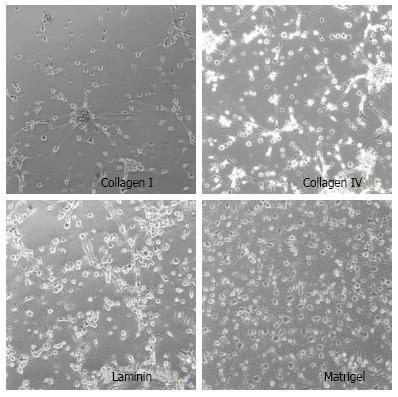
Figure 5 Stem cells cultured on Petri dishes coated with different matrix, showing non-physiologically relevant morphology with a few neurite growth.
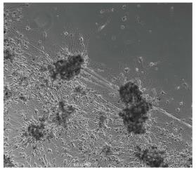
Figure 6 An engineered brain tumor tissue graft in culture, showing tumor lesions (black dots) that attract stem cells to engraft.
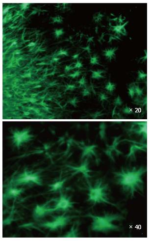
Figure 7 Neural stem cells cultured on the engineered tissue graft showing abundant neurite formation and neuronal morphology.
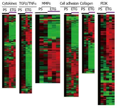
Figure 8 Data analysis of Affymetrix Gene Chip arrays for pediatric derived brain tumor stem cells grown on engineered tissue graftmatrix-like surface or polystyrene dish.
The cells were grown on engineered tissue graft (ETG) matrix-like surface or polystyrene dish (PS) for 7 d for gene chip array analysis showing gene clusters on different functional group of signaling pathways. Notice that red color represents the highest expression, green color for medium expression, and black for lowest expression. MMPs: Matrix-remodeling matrix metalloproteinases; TNF: Tumor necrosis factors; TGF: Transforming growth factor.
Figure 9 An engineered tissue graft is used as the designer matrix to train stem cells to target a specific tumor as shown for production of a morphologically homogeneous population of stem cells.
- Citation: Li SC, Kabeer MH, Vu LT, Keschrumrus V, Yin HZ, Dethlefs BA, Zhong JF, Weiss JH, Loudon WG. Training stem cells for treatment of malignant brain tumors. World J Stem Cells 2014; 6(4): 432-440
- URL: https://www.wjgnet.com/1948-0210/full/v6/i4/432.htm
- DOI: https://dx.doi.org/10.4252/wjsc.v6.i4.432


