修回日期: 2006-03-01
接受日期: 2006-03-07
在线出版日期: 2006-04-28
目的: 探讨C-myc基因蛋白在眼附属器和胃肠道MALT型淋巴瘤的表达和作用.
方法: 应用免疫荧光法检测C-myc在眼附属器淋巴瘤(n = 32)、胃肠道淋巴瘤(n = 29)以及正常眼附属器组织(n = 32)和正常胃肠道淋巴组织(n = 29)中的表达.
结果: C-myc在眼附属器淋巴瘤和胃肠道淋巴瘤中的表达率分别为68.8%和86.2%, 与年龄和性别无关(P>0.05). 胃肠道MALT型淋巴瘤中, 有9例表现为高分期转化病灶, 与眼附属器淋巴瘤(1例)有显著差异(P<0.05). C-myc在正常眼附属器组织和正常胃肠道淋巴组织中无表达.
结论: 眼部附属器MALT型淋巴瘤中C-myc表达率比在胃肠道MALT型淋巴瘤中低, 眼部附属器MALT型淋巴瘤和胃肠道MALT型淋巴瘤预后好.
引文著录: 苏颖, 王峰. 胃肠道和眼附属器MALToma C-myc的表达. 世界华人消化杂志 2006; 14(12): 1219-1221
Revised: March 1, 2006
Accepted: March 7, 2006
Published online: April 28, 2006
AIM: To investigate the expression of C-myc protein in ocular adenxa and gastrointestinal mucosa-associated lymphatic tissue lymphomas (MALToma) and its significance.
METHODS: Immunofluorescence assay was used to detect the expression of C-myc protein in lymphoma of the ocular adnexa (n = 32) and gastrointestinal MALToma (n = 29) as well as their corresponding normal tissues.
RESULTS: The positive rate of C-myc expression was 68.8% and 86.2% in lymphoma of the ocular adnexa and gastrointestinal MALToma, respectively, and C-myc expression had no significant correlations with the age and sex of patients (P > 0.05). In gastrointestinal MALToma, 9 cases were highly-differentiated focus, which was significantly different from that in lymphoma of the ocular adnexa (only 1 case). C-myc was not expressed in normal ocular adenxa and gastrointestinal lymphatic tissues.
CONCLUSION: The expression of C-myc protein is higher in lymphoma of the ocular adnexa, and the patients with ocular adenxa lymphoma and gastrointestinal MALToma have a good prognosis.
- Citation: Su Y, Wang F. Expression of C-myc in ocular adnexa and gastrointestinal mucosa-associated lymphatic tissue lymphomas. Shijie Huaren Xiaohua Zazhi 2006; 14(12): 1219-1221
- URL: https://www.wjgnet.com/1009-3079/full/v14/i12/1219.htm
- DOI: https://dx.doi.org/10.11569/wcjd.v14.i12.1219
在黏膜上皮下的固有层内存在着淋巴细胞集合部位, 形成一定程度上独立于全身免疫机构之外的固有免疫组织,即黏膜相关淋巴组织(mucosa associated lymph tissue, MALT)[1], 担负着防御抗原入侵的任务, 为机体提供较为完善的保护[2-4]. 在眼部附属器如泪腺、泪囊和泪道黏膜下同样存在MALT. 我们观察C-myc基因蛋白在眼部附属器和胃肠道黏膜相关淋巴组织淋巴瘤(MALToma)的表达.
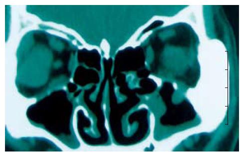
确诊眼部附属器MALToma. 男23例, 女9例. 年龄15-65(平均52)岁. 病灶局限于泪腺25例, CT提示为泪腺肿物(图1), 在眼睑中7例, 通过Ann Arbor临床分期, 31例中Ⅰ期, 局限于眼部附属器1例, Ⅱ期1例. 29例胃肠道MALToma, 男21例, 女8例. 年龄19-63(平均45)岁. 病灶局限于胃部15例, 在肠部14例. 根据Musshoff临床分期标准, 21例为Ⅰ级, 6例为Ⅱ级, 2例为Ⅳ级. 诊断采用WHO标准. 32例确诊眼部附属器MALToma和29例胃肠道MALToma作为实验组. 32例正常眼附属器组织和29例正常胃肠道相关淋巴组织作为对照组. 石蜡包埋组织切片4 μm厚, 经40 g/L甲醛固定, C-myc(1:100)抗体和Texas red抗人IgG为美国Serotec公司提供.
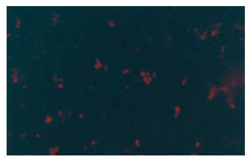
免疫组织化学间接荧光法, 于荧光显微镜下观察. C-myc阳性指标为红色荧光, 用图像分析系统分析.
统计学处理 数据分析应用χ2检验和SPSS10.0软件.
32例眼部附属器MALT型淋巴瘤中, C-myc阳性率为68.8%. C-myc的表达与年龄、性别、部位或临床分期没有显著关系.
在29例胃肠道MALT型淋巴瘤中, C-myc阳性率为86.2%(图2). 通过统计学分析, C-myc表达与年龄、性别和病灶部位没有显著关系. 在32例眼部MALT型淋巴瘤中, 1例为高分期转化病灶. 而在29例胃肠道MALT型淋巴瘤中, 9例表现为高分期转化病灶. 二者之间有显著差异(P<0.05).
在32例正常眼附属器组织和29例正常胃肠道相关淋巴组织中未见C-myc表达.
黏膜相关淋巴组织淋巴瘤(MALToma)是眼部附属器淋巴瘤中最常见的淋巴瘤(80%)[5]. 在临床中, MALToma进展缓慢并且组织病理学等级为Ⅰ[6,7]. 其低度恶性, 很少转移和高度良性转化, 并且通常有较好预后[8]. C-myc基因是髓细胞白血病病毒癌基因的同系物, 由3个外显子和2个内含子组成, 定位于人类染色体8q24, 编码一种p62核蛋白. 在细胞静止期, C-myc几乎不表达, 但在有丝分裂原刺激下迅速表达促使细胞由G0期进入G1期, 增加细胞众数. 因此, C-myc原癌基因与细胞周期调控有关. C-myc原癌基因激活的主要机制有扩增、重排和异常高表达. 前二者为C-myc基因结构异常, 后者则与C-myc基因的调控有关, C-myc基因低甲基化可能为其激活的另一重要机制.
1985年Shibuya首次在部分胃癌细胞中发现C-myc基因扩增和高表达. C-myc表达与肿瘤生长速度和分化程度有关, 在增殖速度较快和分化较低的肿瘤中表达增高更为显著. 我科对35例胃癌进行检测仅1例有扩增, 说明C-myc基因扩增在胃癌中并非常见. 虽然胃癌C-myc扩增率较低(国外报告低于10%), 但C-myc的表达却相对较高. Tatsuta et al采用原位杂交技术检测31例胃黏膜隆起性病变mRNA的表达, 正常胃黏膜均为阴性, 18例腺瘤中有4例(22%)呈弱阳性, 而26例胃癌中有16例(62%)C-myc mRNA呈阳性. 随访结果表明, C-myc mRNA阳性病例在随访过程中有近半数发生癌变, 而mRNA阴性病例未发现有癌变病例. Ninomiye et al采用免疫组化法分析213例胃癌C-myc p62蛋白的高表达与肿瘤的浸润、深度和预后不良有关.
MALToma是一种低分期淋巴瘤[8], 细胞增殖通常低于30%. 当其增殖高于30%时, 容易转移至其他部位. 在32例眼部附属器MALToma中, C-myc阳性率为68.8%. 实验表明眼附属器MALToma预后与临床分期相关, 因为只有2例分别在Ⅰ期和Ⅱ期. 眼部附属器MALToma在不同时期有不同治疗. 在Ⅰ期的病例可通过手术和局部放疗治疗. Ⅱ-Ⅳ病例必须化疗和应用大量放疗. Thieblemont et al[9]研究表明在眼部附属器MALToma和胃肠道MALToma的预后没有显著差异. 我们的研究表明眼部附属器MALToma中C-myc表达率比在胃肠道MALToma中低, 眼部附属器MALToma和胃肠道MALToma预后好. 有许多研究关于胃肠道MALToma, 可以作为眼部附属器MALToma诊断治疗和预后的参考.
本文研究C-myc基因蛋白在眼附属器和胃肠道MALT型淋巴瘤的表达和作用, 立意有一定新颖性, 研究符合伦理学要求.
编辑: 潘伯荣 电编:李琪
| 1. | Kuo SH, Chen LT, Chen CL, Doong SL, Yeh KH, Wu MS, Mao TL, Hsu HC, Wang HP, Lin JT. Expression of CD86 and increased infiltration of NK cells are associated with Helicobacter pylori-dependent state of early stage high-grade gastric MALT lymphoma. World J Gastroenterol. 2005;11:4357-4362. [PubMed] |
| 2. | de Jong D, Boot H, van Heerde P, Hart GA, Taal BG. Histological grading in gastric lymphoma: pretreatment criteria and clinical relevance. Gastroenterology. 1997;112:1466-1474. [PubMed] |
| 3. | Cheng H, Wang J, Zhang CS, Yan PS, Zhang XH, Hu PZ, Ma FC. Clinicopathologic study of mucosa-associated lymphoid tissue lymphoma in gastroscopic biopsy. World J Gastroenterol. 2003;9:1270-1272. [PubMed] |
| 4. | Boot H, de Jong D. Gastric lymphoma: the revolution of the past decade. Scand J Gastroenterol. 2002;27-36. [PubMed] |
| 5. | Morgner A, Miehlke S, Fischbach W, Schmitt W, Muller-Hermelink H, Greiner A, Thiede C, Schetelig J, Neubauer A, Stolte M. Complete remission of primary high-grade B-cell gastric lymphoma after cure of Helicobacter pylori infection. J Clin Oncol. 2001;19:2041-2048. [PubMed] |
| 6. | Lee SL, Lee YC, Chung JB. Low grade gastric MALToma: Treatment strategies based on 10 year follow-up. World J Gastroenterol. 2004;10:223-226. |
| 7. | Nakamura S, Matsumoto T, Suekane H, Takeshita M, Hizawa K, Kawasaki M, Yao T, Tsuneyoshi M, Iida M, Fujishima M. Predictive value of endoscopic ultrasonography for regression of gastric low grade and high grade MALT lymphomas after eradication of Helicobacter pylori. Gut. 2001;48:454-460. [PubMed] |
| 8. | Greiner A, Knorr C, Qin Y. Low-grade B-cell lymphomas of mucosa-associated lymphoid tissue (MALT-type) require CD40-mediated signalling and Th2-type cytokines for in vitro growth and differentiation. Am J Pathol. 1997;150:1583-1593. |
| 9. | Thieblemont C, de la Fouchardiere A, Coiffier B. Nongastric mucosa-associated lymphoid tissue lymphomas. Clin Lymphoma. 2003;3:212-224. [PubMed] |


