Copyright
©2009 The WJG Press and Baishideng.
World J Gastroenterol. May 7, 2009; 15(17): 2162-2165
Published online May 7, 2009. doi: 10.3748/wjg.15.2162
Published online May 7, 2009. doi: 10.3748/wjg.15.2162
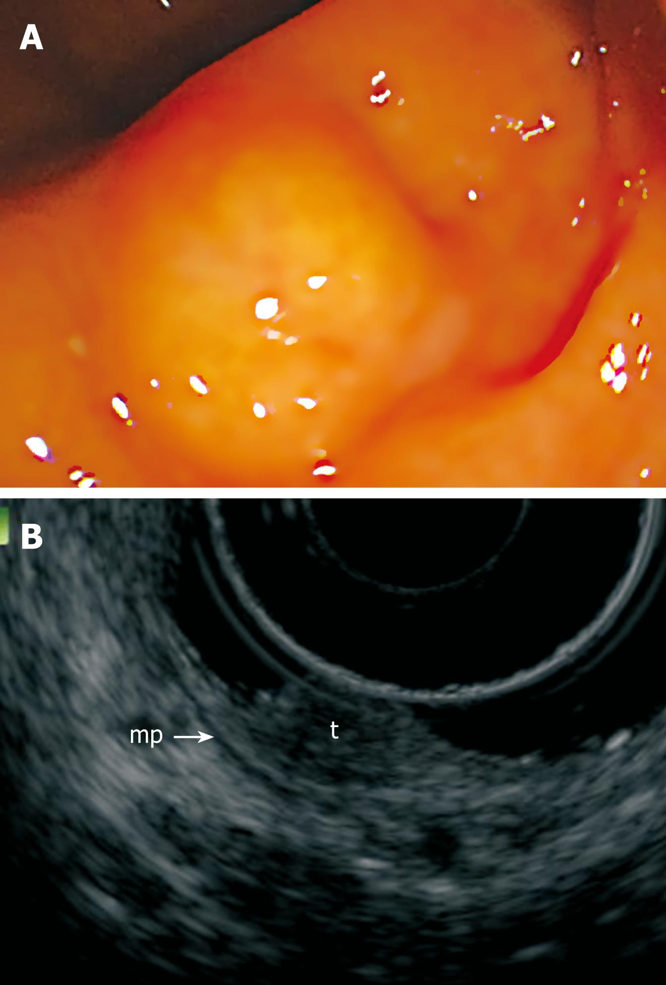
Figure 1 Pretherapeutic examinations of rectal carcinoid.
A: Endoscopic view of the small rectal carcinoid; B: EUS showing a hypoechoic solid tumor (t) in the superficial submucosa. Arrow-mp: Muscularis propria.
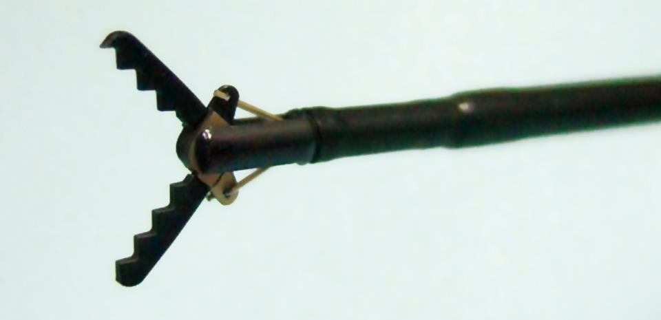
Figure 2 Distal tip of the GSF.
The outer side of the forceps is insulated so that electrosurgical current energy is concentrated at the blade to avoid burning the surrounding tissue.
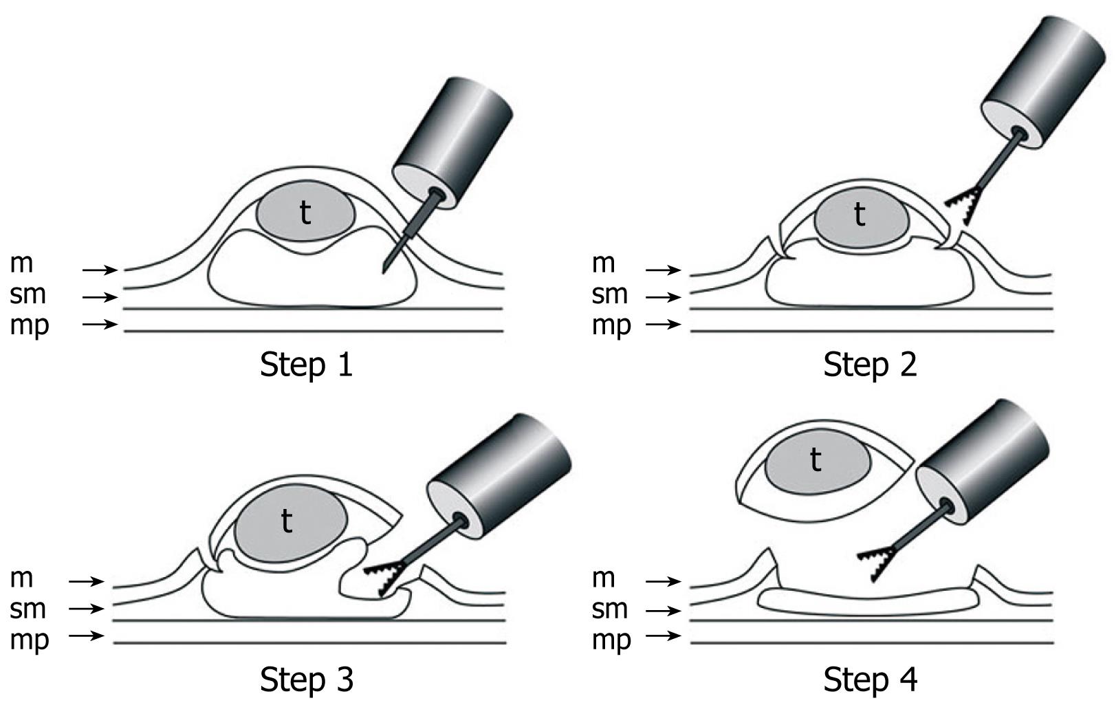
Figure 3 Schematic shows ESD using GSF.
Step 1: A concentrated glycerin solution mixed with a small volume of epinephrine and indigo carmine dye is injected into the submucosal layer around the target lesion to lift the entire lesion; Step 2: The lesion is separated from the surrounding normal mucosa by complete incision around the lesion using the GSF; Step 3: A piece of submucosal tissue is grasped and cut with the forceps using an electrosurgical current to effect submucosal exfoliation; Step 4: The lesion is resected in one piece. m: Mucosa; sm: Submucosa; mp: Muscularis propria; t: Tumor.
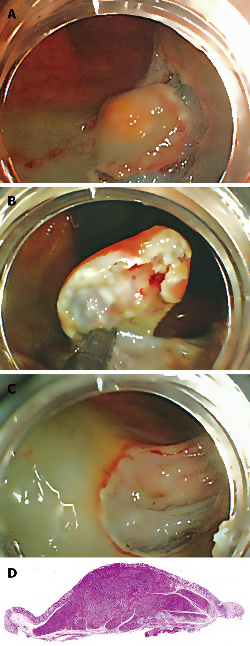
Figure 4 ESD using GSF of rectal carcinoid.
A: Endoscopic view of the partial circumferential incision of the tumor using GSF; B: Endoscopic view of the submucosal exfoliation under the tumor using GSF; C: The lesion is cut completely from the muscle layer; D: The resected specimen showing curative en bloc resection of the lesion.
- Citation: Akahoshi K, Motomura Y, Kubokawa M, Matsui N, Oda M, Okamoto R, Endo S, Higuchi N, Kashiwabara Y, Oya M, Akahane H, Akiba H. Endoscopic submucosal dissection of a rectal carcinoid tumor using grasping type scissors forceps. World J Gastroenterol 2009; 15(17): 2162-2165
- URL: https://www.wjgnet.com/1007-9327/full/v15/i17/2162.htm
- DOI: https://dx.doi.org/10.3748/wjg.15.2162


